Skip to content
Medical Image Analysis Laboratory

Medical Image Analysis Laboratory

Follow us

  • LinkedIn
  • Twitter

Menu

  • About us
  • Team
    • Meritxell Bach Cuadra
    • Hélène Lajous
    • João Jorge
    • Thomas Sanchez
    • Yasser Alemán-Gómez
    • Andrés le Boeuf Fló
    • Cristina Sainz Martinez
    • Hamza Kebiri
    • Jaime Barranco
    • Joe Najm
    • Nataliia Molchanova
    • Maxence Wynen
    • Miguel Díaz-Cacho Dafonte
    • Priscille de Dumast
    • Samuel Lamon
    • Tommaso Di Noto
    • Alumni
  • Research
    • Projects
      • Super-Resolution T2w Foetal Brain MRI for Computer Assisted Diagnosis and Quantitative Analysis
      • Super-resolution reconstruction of in vivo brain diffusion MRI
      • Quantitative MR Imaging (qMRI) of the in vivo fetal brain
      • MSxplain
      • Machine learning methods for 3T & 7T MRI analysis of white matter and cortical lesions in Multiple Sclerosis
      • Structural analysis of the thalamic nuclei in early psychosis and schizophrenia
      • Targeting the venture-intermediate thalamic nucleus at 3T and 7T MRI
    • Publications
  • Open data & software
  • Teaching
    • Courses and Seminars
    • Student projects
  • News
    • Highlights
    • Fun
    • Calendar
  • Contact

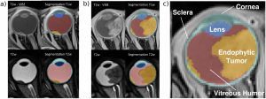
Multi-channel MRI segmentation of eye structures and tumors

Meritxell Bach Cuadra Software 24 March 202024 March 2020

This is an open source software for computing the automatic segmentation of eye structures and tumors in 3D Magnetic Resonance Imaging. Read more …

  • ← MBIS: Multivariate Bayesian Image Segmentation tool
  • 3T and 7T MRI brain images: targeting the Ventro-intermediate thalamic nucleus →
Copyright © 2023 Medical Image Analysis Laboratory. All rights reserved.
Theme: Accelerate by ThemeGrill. Powered by WordPress.
  • Home
  • Download
  • Blog
  • Layout
  • Sample Page