
Easy walk from Chalet-à-Gobet till Lausanne, April 2022
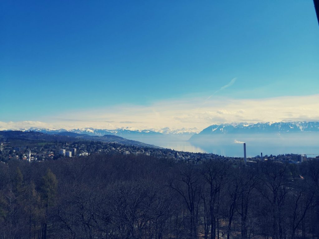
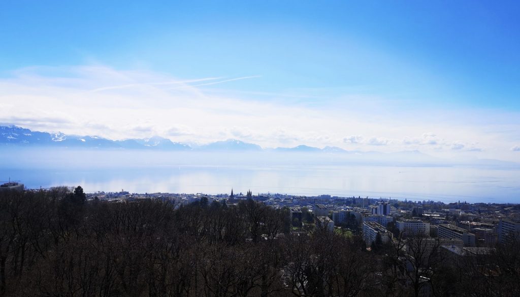
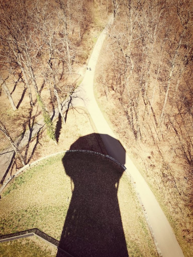
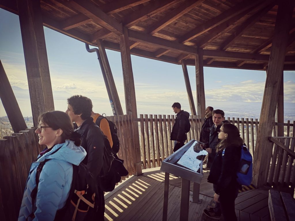
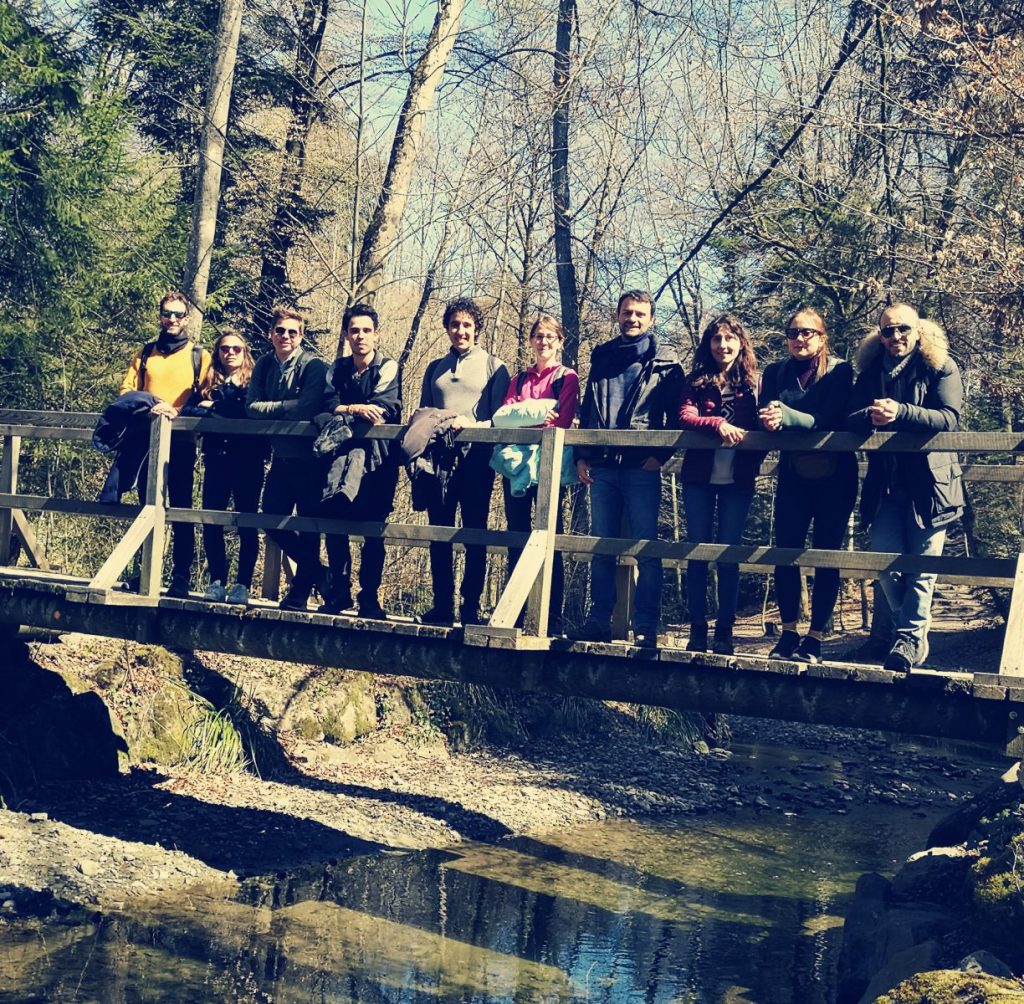
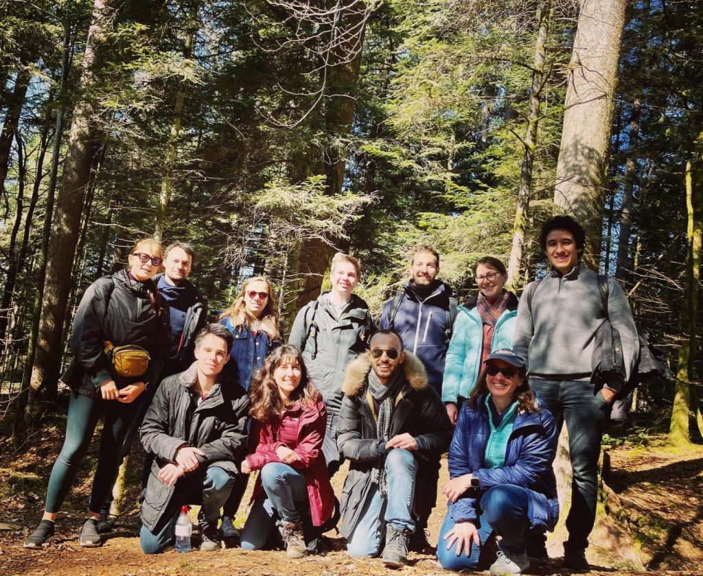
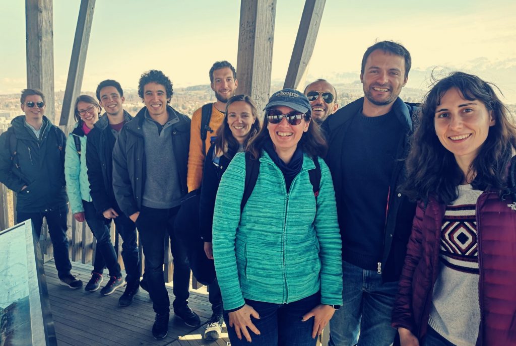
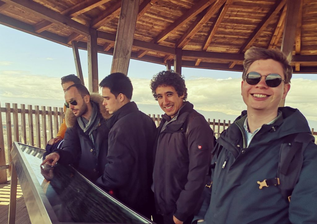
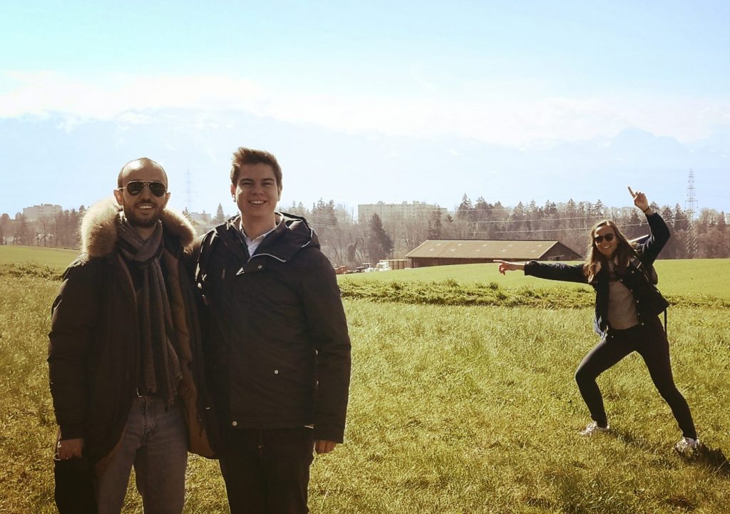
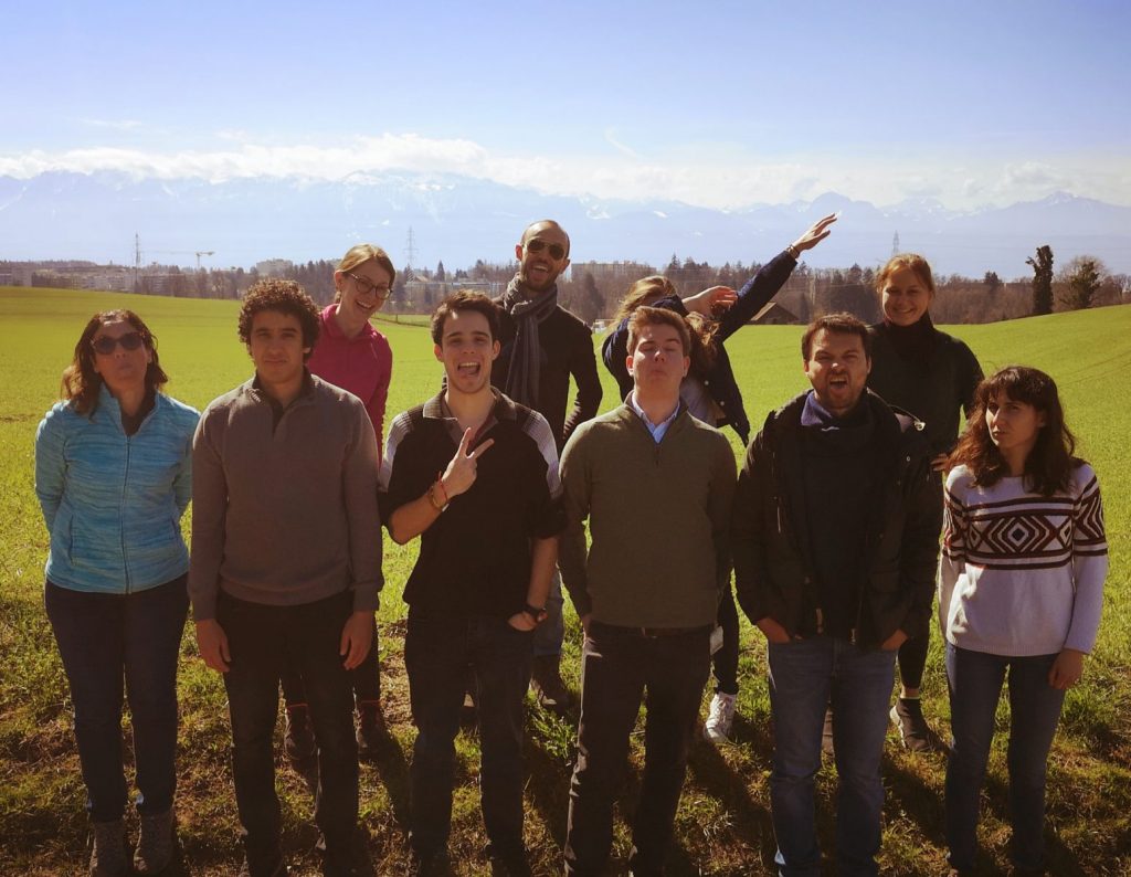
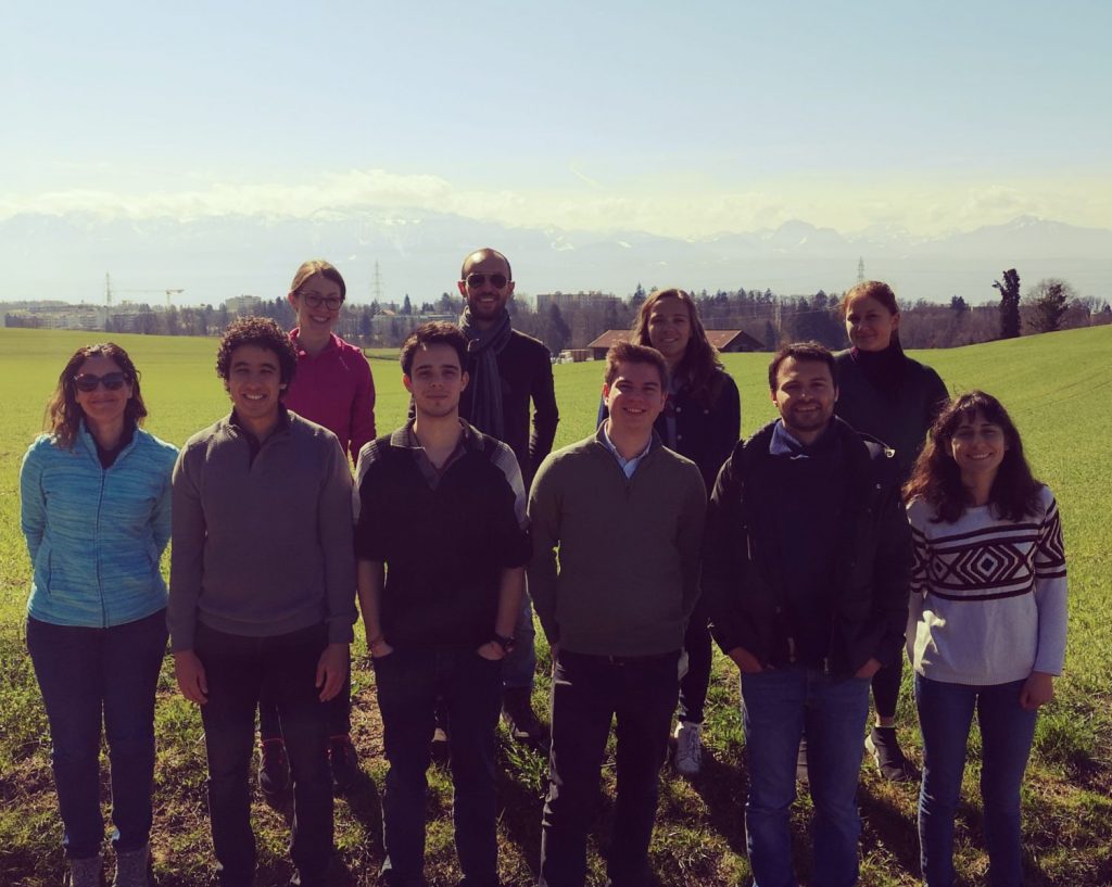
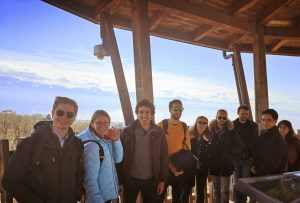
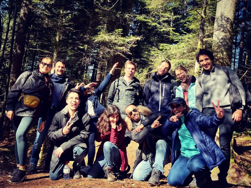

Easy walk from Chalet-à-Gobet till Lausanne, April 2022
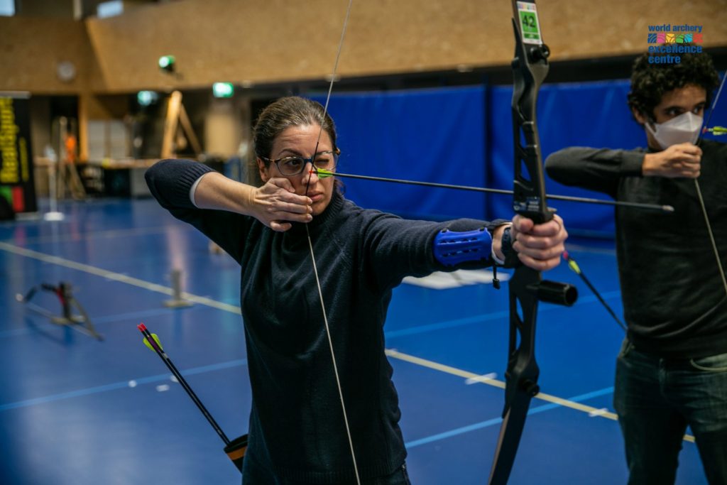
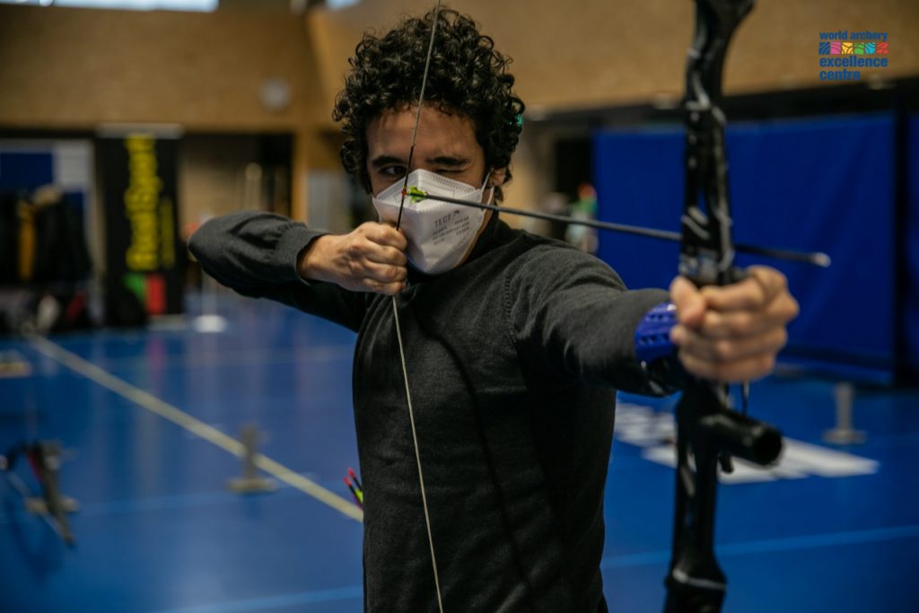
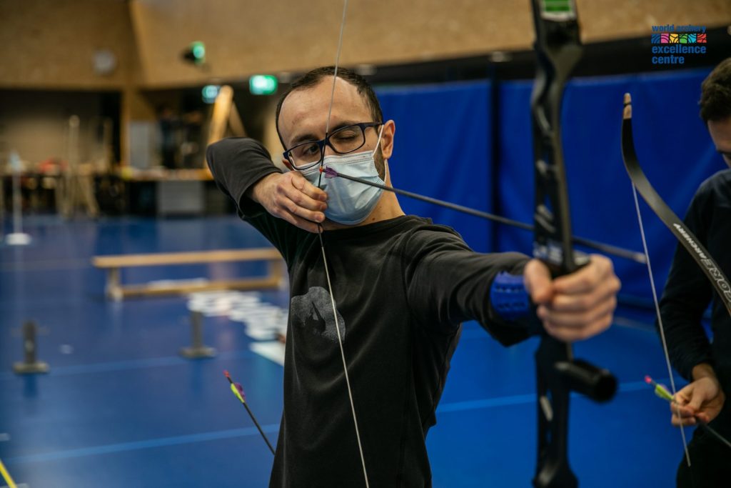
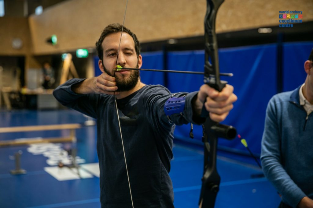
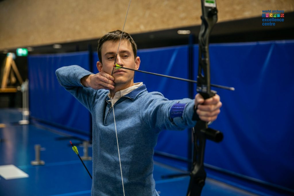
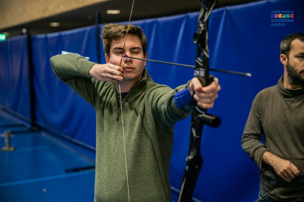
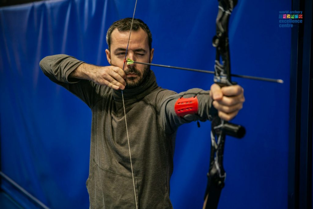
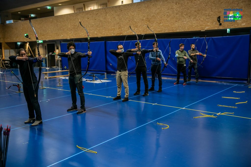
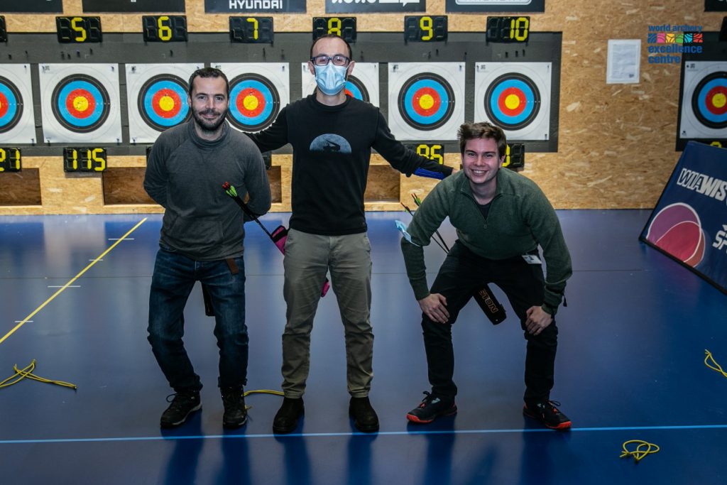
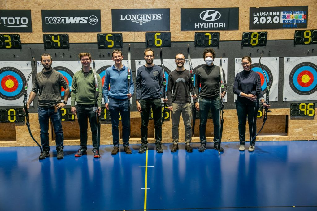
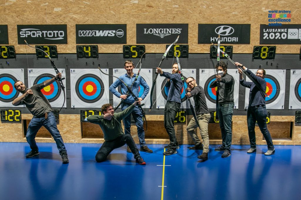
World Archery Excellence Centre (December 2021Lausanne, Switzerland)
Watch the seminar by H. Lajous on “T2 quantification from Super-resolution reconstructed Clinical Fast Spin Echo MR Acquisitions”, part of this seminar shows our recent work accepted to MICCAI 2020.
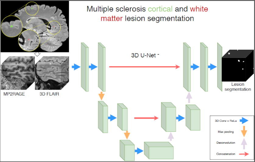
New accepted paper in NeuroImage: Clinical by F. La Rosa and co-authors on automated cortical and white matter lesions in multiple sclerosis at 3T.
We have accepted two paper for MICCAI 2020 !! Keep posted to know more about 7T MRI segmentation of cortical multiple sclerosis lesions and Super-resolution T2 mapping from clinical fast spin echo MRI.
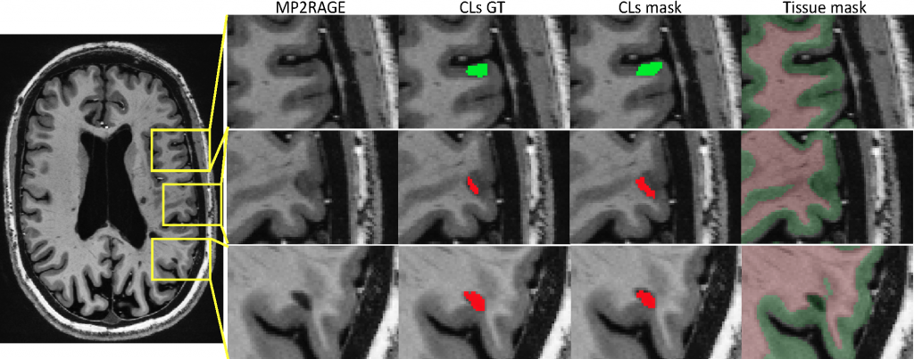
Automated Detection of Cortical Lesions in Multiple Sclerosis Patients with 7T MRI
F. La Rosa 1,2,3, Erin S Beck 4, A. Abdulkadir 5,6, J.-Ph. Thiran 1,3, D. S Reich 4, P. Sati 4,7, and M. Bach Cuadra 3,2,1
1 LTS5, Ecole Polytechnique Federale de Lausanne, Switzerland
2 CIBM, University of Lausanne, Switzerland
3 Radiology Department, Lausanne University Hospital, Switzerland
4 Translational Neuroradiology Section, NINDS, NIH, Bethesda, MD, USA
5 Universitare Psychiatrische Dienste and University of Bern, Switzerland
6 CBICA, University of Pennsylvania, Philadelphia, USA
7 Department of Neurology, Cedars-Sinai Medical Center, Los Angeles, CA, USA
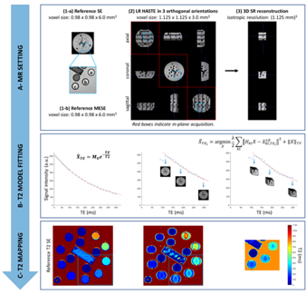
T2 Mapping from Super-Resolution-Reconstructed Clinical Fast Spin Echo Magnetic Resonance Acquisitions
H. Lajous 1,2, T. Hilbert 1,3,4, C. W. Roy 1, S. Tourbier 1, P. de Dumast 1,2, T. Yu 4, J.-Ph. Thiran 1,4, J.-B. Ledoux 1,2, D. Piccini 1,3, P. Hagmann 1, R. Meuli 1, T. Kober 1,3,4, M. Stuber 1,2, R.B. van Heeswijk 1, M. Bach Cuadra 1,2,4
1 Department of Radiology, Lausanne University Hospital (CHUV) and University of Lausanne (UNIL), Switzerland
2 Center for Biomedical Imaging (CIBM), Lausanne, Switzerland
3 Advanced Clinical Imaging Technology (ACIT), Siemens Healthcare, Switzerland
4 LTS5, Ecole Polytechnique Federale de Lausanne
New paper accepted in “NMR in Biomedicine” by Pietro Maggi an co-authors on automated central vein sign assessment in multiple sclerosis.
New paper accepted in Magnetic Resonance in Medicine Imaging by João Jorge and co-authors related to several methodological improvements to enhance 7T SWI quality and intensity contrast, specifically for an improved visualisation of the human thalamus.
New published paper in “Seminars in Musculoskeletal Radiology” reviewing Machine Learning Segmentation and Radiomics in Musculoskeletal Imaging. This work is in collaboration with J. Favre and P. Omoumi (CHUV).
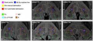 We provide 3T and 7T human brain Magnetic Resonance Imaging for the automated segmentation and functional neurosurgery targeting of the Ventro-intermediate thalamic nucleus. Read more …
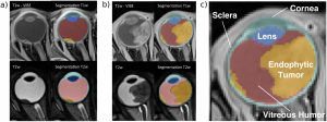
This is an open source software for computing the automatic segmentation of eye structures and tumors in 3D Magnetic Resonance Imaging. Read more …