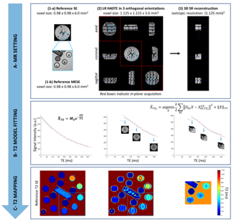
Watch the seminar by H. Lajous on “T2 quantification from Super-resolution reconstructed Clinical Fast Spin Echo MR Acquisitions”, part of this seminar shows our recent work accepted to MICCAI 2020.
MRI
MICCAI 2020 papers: congratulations to H. Lajous and F. La Rosa !
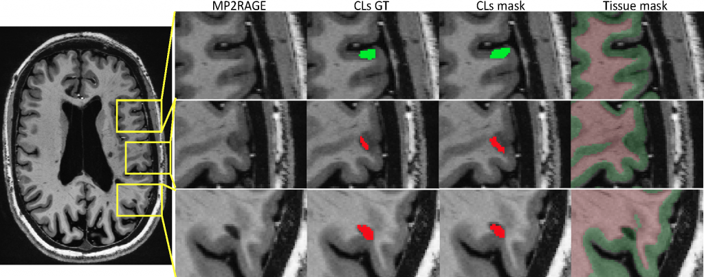
We have accepted two paper for MICCAI 2020 !! Keep posted to know more about 7T MRI segmentation of cortical multiple sclerosis lesions and Super-resolution T2 mapping from clinical fast spin echo MRI.

Automated Detection of Cortical Lesions in Multiple Sclerosis Patients with 7T MRI
F. La Rosa 1,2,3, Erin S Beck 4, A. Abdulkadir 5,6, J.-Ph. Thiran 1,3, D. S Reich 4, P. Sati 4,7, and M. Bach Cuadra 3,2,1
1 LTS5, Ecole Polytechnique Federale de Lausanne, Switzerland
2 CIBM, University of Lausanne, Switzerland
3 Radiology Department, Lausanne University Hospital, Switzerland
4 Translational Neuroradiology Section, NINDS, NIH, Bethesda, MD, USA
5 Universitare Psychiatrische Dienste and University of Bern, Switzerland
6 CBICA, University of Pennsylvania, Philadelphia, USA
7 Department of Neurology, Cedars-Sinai Medical Center, Los Angeles, CA, USA

T2 Mapping from Super-Resolution-Reconstructed Clinical Fast Spin Echo Magnetic Resonance Acquisitions
H. Lajous 1,2, T. Hilbert 1,3,4, C. W. Roy 1, S. Tourbier 1, P. de Dumast 1,2, T. Yu 4, J.-Ph. Thiran 1,4, J.-B. Ledoux 1,2, D. Piccini 1,3, P. Hagmann 1, R. Meuli 1, T. Kober 1,3,4, M. Stuber 1,2, R.B. van Heeswijk 1, M. Bach Cuadra 1,2,4
1 Department of Radiology, Lausanne University Hospital (CHUV) and University of Lausanne (UNIL), Switzerland
2 Center for Biomedical Imaging (CIBM), Lausanne, Switzerland
3 Advanced Clinical Imaging Technology (ACIT), Siemens Healthcare, Switzerland
4 LTS5, Ecole Polytechnique Federale de Lausanne
New paper: CVSnet – A machine learning approach for automated central vein sign assessment in multiple sclerosis
New paper accepted in “NMR in Biomedicine” by Pietro Maggi an co-authors on automated central vein sign assessment in multiple sclerosis.
New paper: Improved susceptibility‐weighted imaging for high contrast and resolution thalamic nuclei mapping at 7T
New paper accepted in Magnetic Resonance in Medicine Imaging by João Jorge and co-authors related to several methodological improvements to enhance 7T SWI quality and intensity contrast, specifically for an improved visualisation of the human thalamus.
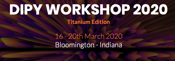
DIPY workshop, Bloomington – Indiana, USA, 2020
 Hamza Kebiri will participate at the DIPY workshop, Bloomington – Indiana, USA, 2020 and he will present the foetal diffusion MR image super-resolution reconstruction we are developing in our FNS project. It will be a great opportunity to disseminate our ongoing research among international experts in the brain diffusion MRI community !
MIALSRTK: Super-resolution reconstruction of fetal MRI


The Medical Image Analysis Laboratory Super-resolution toolkit (MIALSRTK) consists of a set of C++ image processing tools necessary to perform motion-robust super-resolution foetal MRI reconstruction. Read more…
