Watch the seminar by H. Lajous on “T2 quantification from Super-resolution reconstructed Clinical Fast Spin Echo MR Acquisitions”, part of this seminar shows our recent work accepted to MICCAI 2020.
Work news
New paper: Multiple sclerosis cortical and WM lesion segmentation at 3T MRI: a deep learning method based on FLAIR and MP2RAGE
New accepted paper in NeuroImage: Clinical by F. La Rosa and co-authors on automated cortical and white matter lesions in multiple sclerosis at 3T.
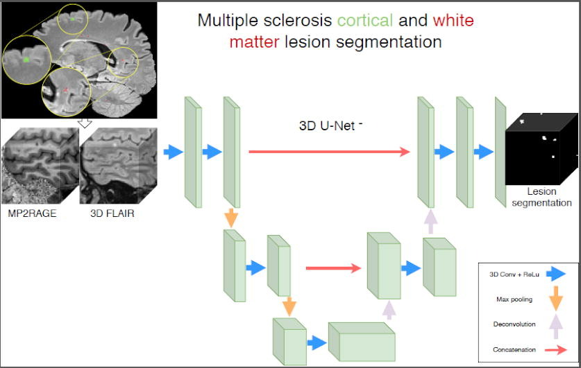
MICCAI 2020 papers: congratulations to H. Lajous and F. La Rosa !
We have accepted two paper for MICCAI 2020 !! Keep posted to know more about 7T MRI segmentation of cortical multiple sclerosis lesions and Super-resolution T2 mapping from clinical fast spin echo MRI.
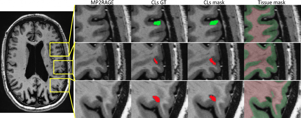
Automated Detection of Cortical Lesions in Multiple Sclerosis Patients with 7T MRI
F. La Rosa 1,2,3, Erin S Beck 4, A. Abdulkadir 5,6, J.-Ph. Thiran 1,3, D. S Reich 4, P. Sati 4,7, and M. Bach Cuadra 3,2,1
1 LTS5, Ecole Polytechnique Federale de Lausanne, Switzerland
2 CIBM, University of Lausanne, Switzerland
3 Radiology Department, Lausanne University Hospital, Switzerland
4 Translational Neuroradiology Section, NINDS, NIH, Bethesda, MD, USA
5 Universitare Psychiatrische Dienste and University of Bern, Switzerland
6 CBICA, University of Pennsylvania, Philadelphia, USA
7 Department of Neurology, Cedars-Sinai Medical Center, Los Angeles, CA, USA
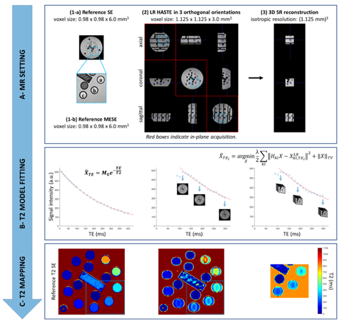
T2 Mapping from Super-Resolution-Reconstructed Clinical Fast Spin Echo Magnetic Resonance Acquisitions
H. Lajous 1,2, T. Hilbert 1,3,4, C. W. Roy 1, S. Tourbier 1, P. de Dumast 1,2, T. Yu 4, J.-Ph. Thiran 1,4, J.-B. Ledoux 1,2, D. Piccini 1,3, P. Hagmann 1, R. Meuli 1, T. Kober 1,3,4, M. Stuber 1,2, R.B. van Heeswijk 1, M. Bach Cuadra 1,2,4
1 Department of Radiology, Lausanne University Hospital (CHUV) and University of Lausanne (UNIL), Switzerland
2 Center for Biomedical Imaging (CIBM), Lausanne, Switzerland
3 Advanced Clinical Imaging Technology (ACIT), Siemens Healthcare, Switzerland
4 LTS5, Ecole Polytechnique Federale de Lausanne
New paper: CVSnet – A machine learning approach for automated central vein sign assessment in multiple sclerosis
New paper accepted in “NMR in Biomedicine” by Pietro Maggi an co-authors on automated central vein sign assessment in multiple sclerosis.
New paper: Improved susceptibility‐weighted imaging for high contrast and resolution thalamic nuclei mapping at 7T
New paper accepted in Magnetic Resonance in Medicine Imaging by João Jorge and co-authors related to several methodological improvements to enhance 7T SWI quality and intensity contrast, specifically for an improved visualisation of the human thalamus.
New paper: Quantification in Musculoskeletal Imaging Using Computational Analysis and Machine Learning: Segmentation and Radiomics
New published paper in “Seminars in Musculoskeletal Radiology” reviewing Machine Learning Segmentation and Radiomics in Musculoskeletal Imaging. This work is in collaboration with J. Favre and P. Omoumi (CHUV).
DIPY workshop, Bloomington – Indiana, USA, 2020
 Hamza Kebiri will participate at the DIPY workshop, Bloomington – Indiana, USA, 2020 and he will present the foetal diffusion MR image super-resolution reconstruction we are developing in our FNS project. It will be a great opportunity to disseminate our ongoing research among international experts in the brain diffusion MRI community !
European Congress of MR in Neuropediatrics ECMRN 2020, Marseille, France
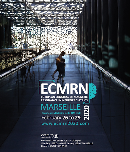 MIAL will participate with three posters at the European Congress of Magnetic Resonance in Neuropediatrics (ECMRN) that will be held in Marseille from 26 to 29th February ! Amazing pre-congress day including foetal MRI… check their program here.
TRABIT summer school
In the context of the European ITN Translational Brain Imaging Network (TRABIT) we organise a Computational Magnetic Resonance Brain Imaging Summer School at EPFL, June 24-28 2019, EPFL, Lausanne.
More information https://trabit2019.epfl.ch
