
Master & bachelor projects
Mapping functional connectivity in callosal dysgenesis
PROJECT DESCRIPTION: The corpus callosum is the largest white matter structure in the human brain, containing over 190 million axons that connect the left and right hemispheres. This brain structure is critical for transfer and integration of information throughout the brain implicated in sensory, motor, visuo-motor integration, as well as higher cognitive functions Congenital anomalies of the corpus callosum, known as callosal dysgenesis, is one the most common congenital brain malformation. Callosal dysgenesis is characterised by incomplete or abnormal formation of the corpus callosum and is associated with variable cognitive, behavioural, and neurological outcomes (e.g., Siffredi et al., 2021). A recently developed neuroimaging method, named apparent diffusion coefficient functional MRI (ADC-fMRI), allows to detect neuronal and axonal morphological fluctuations during firing and actional potential transmission (de Riedmatten et al., 2025; Spencer et al., 2025). This approach provides a robust functional connectivity mapping across both grey and white matter. The application of ADC-fMRI to a clinical population with specific alteration of white-matter pathways, such as individuals with callosal dysgenesis, could bring significant insight into compensation mechanisms and plastic response occurring in such atypical brain, including how interhemispheric transfer is maintained in the absence of the major commissural pathway.
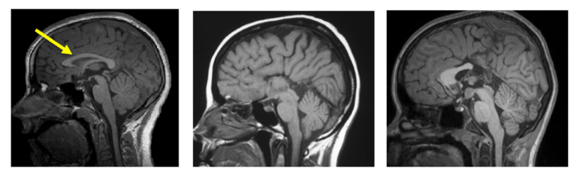
The goal of the project is to analyse ADC-fMRI data from a unique cohort of children with callosal dysgenesis (Schmidt et al., 2026) – from preprocessing of diffusion MRI timeseries to analyses of group differences in brain activity and functional connectivity. Extensive neurobehavioral and clinical assessments are also available, as well as other neuroimaging sequences (such as BOLD fMRI).
This project will be supervised by Dr. Vanessa Siffredi and Prof. Ileana Jelescu. The project bridges neuroscience, child development, signal processing and MRI physics. We are a highly interdisciplinary and collaborative environment within the Radiology Research Unit of the CHUV.
START DATE: Fall 2026 onward
PROFILE: We are seeking a highly motivated master student with interest in pediatric and neuroimaging, and with a background in Electrical/Micro/Life Science Engineering, Neuroscience or Physics. Very good coding skills are essential (Python, bash). Familiarity with neuroimaging software tools is a plus.
INTERESTED? Contact vanessa.siffredi@chuv.ch or ileana.jelescu@chuv.ch.
Microstructural Diffusion MRI in the Rodent Brain
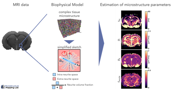
PROJECT DESCRIPTION: Diffusion MRI is a powerful tool to quantify biological tissue microstructure in vivo and non-invasively. The goal of this project is to use advanced biophysical models of diffusion MRI to extract quantitative maps of brain microstructure in animal models of disease vs healthy controls. Possible applications include Parkinson’s disease, creatine deficiency or stroke. Because the estimation of these model parameters can be challenging (degeneracy or high uncertainty), the optimization of model fitting (e.g. via soft constraints or Bayesian uncertainty estimation) to improve biological interpretability is also a possible project lead.
This project will be carried out within the Radiology Research Unit of the CHUV, supervised by Dr. Rita Oliveira and Prof. Ileana Jelescu.
START DATE: January 2026 onward
PROFILE: We are seeking a highly motivated master’s student with a background in Electrical/Micro/Life Science Engineering or Physics. The project is flexible and can be adapted based on the student’s background and on the University programme’s requirements. By the end, you will be able to confidently process neuroimaging data and work with biophysical models of brain microstructure. Very good coding skills are essential.
INTERESTED? Contact Ana.Veiga-De-Oliveira@chuv.ch or ileana.jelescu@chuv.ch.
Unraveling the contrast mechanism of neuromelanin-sensitive MRI
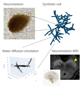
PROJECT DESCRIPTION: Neuromelanin‑sensitive MRI is a novel technique that holds high clinical potential, as it detects pathological alterations in a wide range of diseases, among them Parkinson’s disease and schizophrenia. While its name suggests that it measures the pigment neuromelanin, a surrogate of catecholaminergic function, the physical mechanisms underlying this MRI contrast are debated in the literature. Some researchers even doubt the sequence’s sensitivity to neuromelanin at all. Understanding the underlying physics is essential for quantitative neuromelanin imaging, which could improve early diagnosis and monitoring of neurodegenerative disorders. We aim to explain the neuromelanin-sensitive MRI contrast using computer simulations of the MRI signal. In our lab, we generate synthetic tissue using computer algorithms to study the signal in diffusion MRI. The goal of this master’s project is to extend our computer simulations to include two effects pivotal for the neuromelanin‑sensitive MRI contrast: longitudinal relaxation and magnetization transfer.
This project will be carried out within the Radiology Research Unit of the CHUV, supervised by Dr. Malte Brammerloh and Prof. Ileana Jelescu.
What the student will deliver:
- Implement longitudinal‑relaxation and magnetization‑transfer modules in the existing simulation framework.
- Validate the extended model against published data.
Who we are looking for:
- Strong programming skills (C++)
- Experience with numerical solvers or Monte‑Carlo simulations.
- Basic knowledge of MRI physics (helpful but not mandatory).
- High motivation to model physical processes and interest in neuro‑imaging.
Why join us: The project bridges physics, computer science, and neuroscience, offering an interdisciplinary environment for students eager to contribute to cutting‑edge MRI research.
If you are ready to advance neuromelanin imaging and gain hands‑on experience with state‑of‑the‑art simulation tools, we invite you to apply.
START DATE: January 2026 onward
INTERESTED? Contact malte.brammerloh@chuv.ch or ileana.jelescu@chuv.ch.
Cortical Microstructure Changes in Schizophrenia
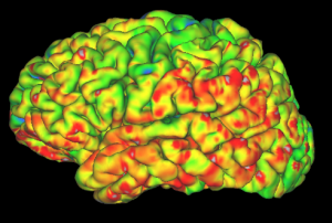
PROJECT DESCRIPTION: Schizophrenia is a psychiatric disease associated with subtle but widespread brain changes. Diffusion MRI is a powerful tool to quantify biological tissue microstructure in vivo and non-invasively. This master project offers a unique opportunity to contribute to translational research in psychiatry and neuroscience. We propose to (A) characterise brain cortical microstructure changes in patients with schizophrenia measured via novel Diffusion MRI markers and (B) relate them to metabolic changes measured using MR Spectroscopy.
This project will be carried out within the Radiology Research Unit of the CHUV, supervised by Tommaso Pavan and Prof. Ileana Jelescu, in collaboration with Prof. Lijing Xin (EPFL).
START DATE: Fall/Winter 2024
PROFILE: We are seeking a highly motivated master student with a background in Electrical, Micro- and Life Science Engineering or Physics. Flexible duration aligned with your University and degree requirements from a research project.
You will learn: to process neuroimaging data, to operate a high-performance computing (HPC) cluster, refine your machine-learning/statistics skills and make a first step into the field of computational psychiatry. Good coding skills are necessary.
INTERESTED? Contact tommaso.pavan@chuv.ch or ileana.jelescu@chuv.ch.