Combo microCT/OI (Xray + Biolum/fluo)- Advanced training

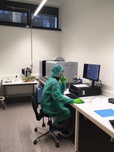
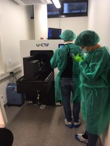
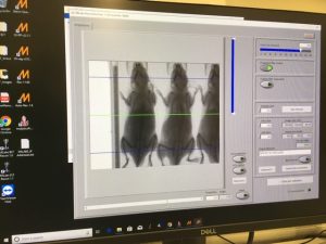
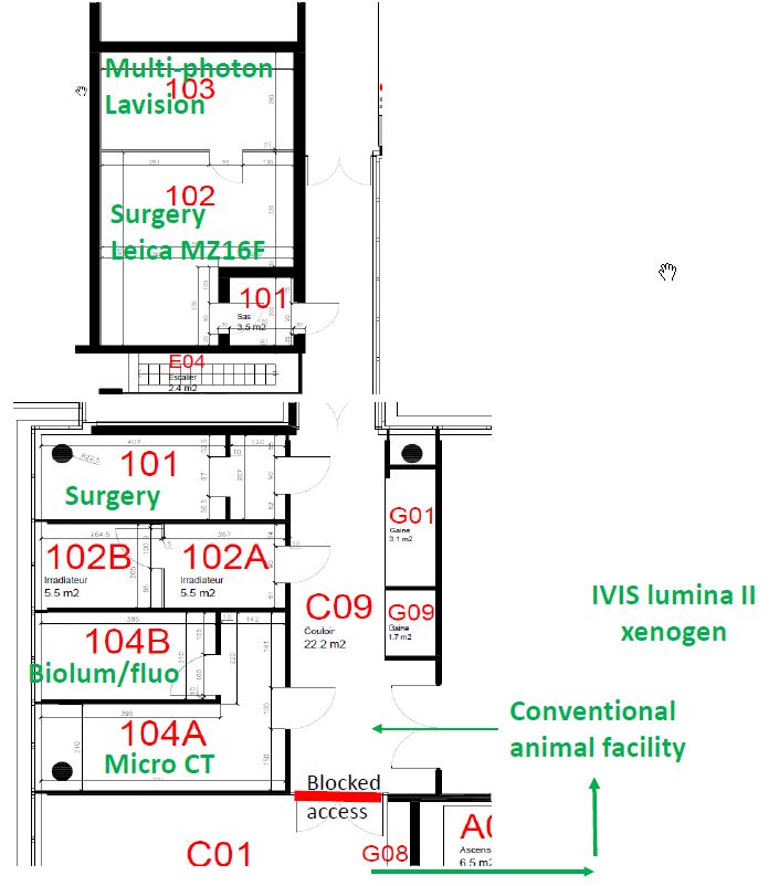
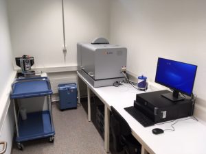
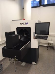
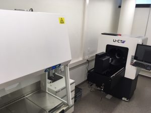
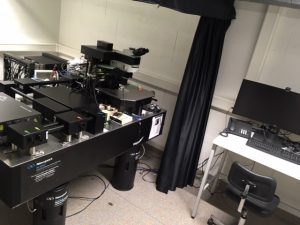
Thanks to all the users participating to the advanced training on the combo optical imaging (Biolum/fluo) and microCT installed at the conventional animal facility CLE C. It was very instructive as it was very diverse, going from Fat pad in vivo imaging to lung nodules, bladder, Caecum and bones. We got a press release after that The University of Lausanne installs a high-end MILabs’ Duet Optical Imager in its core In-Vivo Imaging Laboratory


CLE – Training microCT and Optical Imaging (MILabs)

On Monday, February 10th and Tuesday, February 11th an application specialist from MILabs will be presenting our Hybrid microCT and optical imaging tomograph. This brand-new system will be available to all researchers working in Epalinges.

A public seminar has been organized, as well as specific hands-on training. The latter, however, will be limited to five people, so please contact us  asap if you would like to attend.

Below you can find the program timeline:

Day1 Comments
8:45 9:00 Arrival
9:30 12:00 System demo Depending on the amount of people fitting in the room; otherwise we split the attendees in groups
12:15

13:15

Seminar room B301

Highlighting the capabilities of MILabs’ systems

Covering 2D OI, 3D/CT, CT standalone

A couple of slides about different imaging modalities (SPECT/PET)

For bigger audience

13:15 14:00 Lunch
14:15 17:00 Training presentation

Covering different modules, some precautions, data acquisition, data reconstruction and analysis

For users

Day2
8:45 9:00 Arrival
9:00 12:00 Animal acquisition, BLT/FLT/CT acquisitions, default protocols

Depending on research needs different animal modals preferably with CT and OI contrast

Maximum number of people should be 5

12:00 12:30 Lunch
12:30 14:00 Animal scans reconstruction; manual recon (CT) and advance recon (CT)
14:00 17:00 Data inspection, visualization and analyses


CLE – finalizing installation

Here is an update on installations of the IVIF at the CLE C/D, please let us know if you want to visit: Please be aware that the access to the facility starting from the 18th of November will be through the animal conventional facility (green coat required).

  • Room CLE C 104 has been equipped with a hood for mouse handling, a microCT and a Biolum/fluo (“IVIS like”) detection system from MILabs. Both can be used separately or combined for 3D simultaneous Biolum/Fluo + CT. The IVIS lumina II (xenogen) will be moving to this location ultimately.

  • Room CLE C 102 has been dedicated and totally secured for irradiation with the already exciting Cesium irradiator and the RS2000 for whole cage irradiation.

  • Room CLE C 101 with its stereomicroscope leica M60 is equipped for animal surgery.

  • Room CLE D 102 & 103  intravital microscopy rooms. the first room has been installed with a surgery bench and a Leica stereomicroscope MZ16F while the 103 is hosting the multi-photon microscope from LAVISION.

  • Room CLE C 301 is where you can find IVIF staff and 3 powerful workstations for analyses on dedicated software (Imaris Cell packeage, Living Image 3.2 and 4.7 from perkin elmer, Imalytics from MIlabs…).

CLE – Workstation living image for xenogen IVIS

We have moved the workstation installed with living image 3.2 used for analyzing images previously acquired by the xenogen IVIS. It passed from the CIF to the IVIF in the room CLE C 301. The room can be accessed by a badge with the regular “scientifique” access from the CHUV.

It can be booked on OPEN IRIS  –> scheduler –> In vivo imaging facility CLE –> Workstation IVIF 4 for Biolum/fluo.

Soon we will upgrade to living image 4.7.

thank you for your understanding

CLE – Installation MILabs system final schedule

Here is the updated schedule for the MIlabs installation.

– Tuesday Oct 15th
Unloading, Unpacking, Inspection of the system, System Placement, Power up.

Wednesday Oct 16th
Mechanical checks, Mechanical Calibrations, CT start up.

Thursday Oct 17th
CT Mechanical Checks, CT safety.

Friday Oct 18th
CT Quality Assurance Scans, Emission Detector Calibration.

-Monday Oct 21st
Optical Installation and Calibration.

Tuesday Oct 22nd
Optical Quality Assurance.

Wednesday Oct 23rd & Thursday Oct 24th
User training and Customer Acceptance.