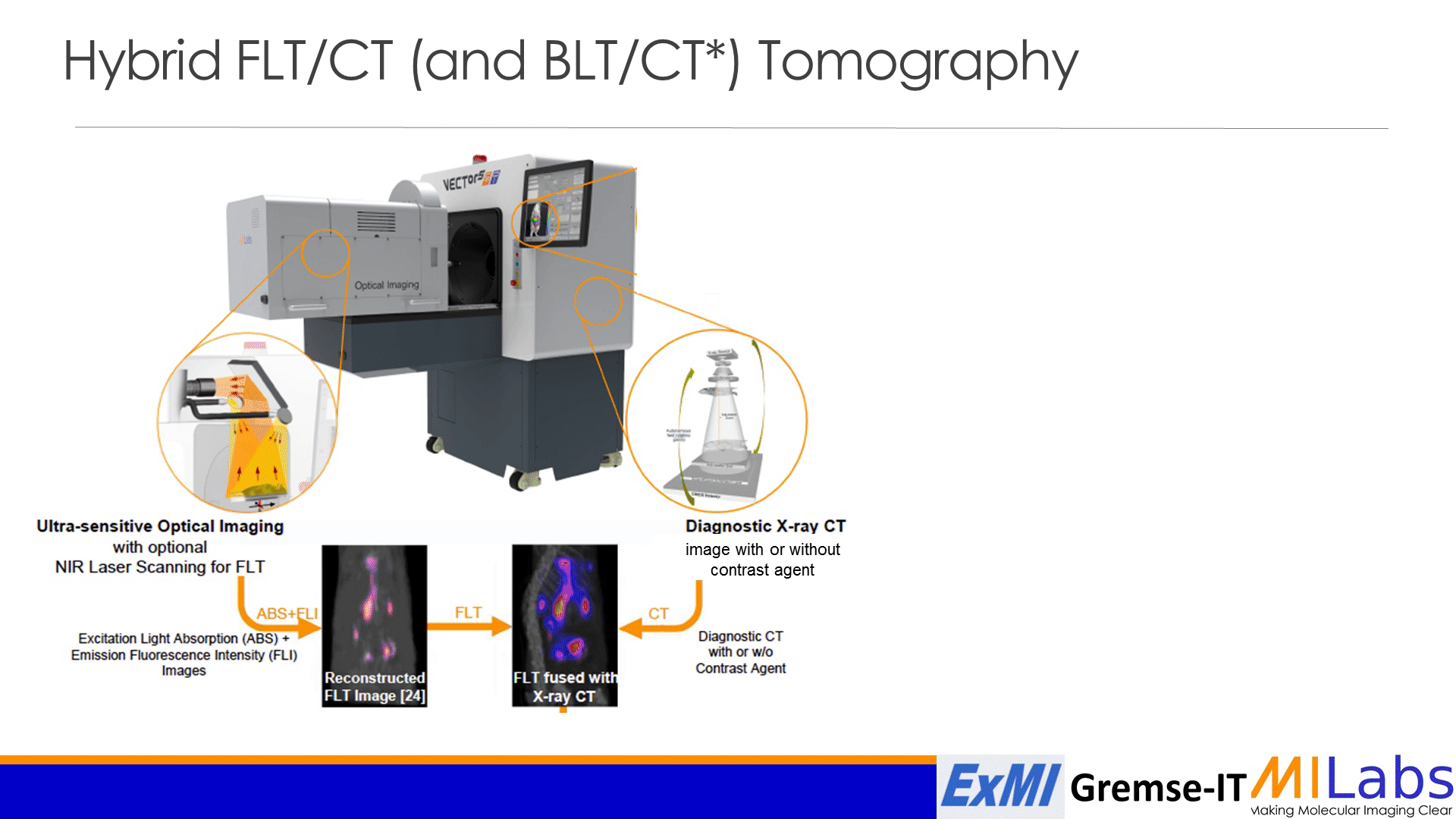
The facility is equipped with a full Imaris version (package for cell biologist, soon release of the 9.8 version) and it is very powerful for analyses of time lapses generated in vivo on our facility.
It is also a useful seminar for all researchers doing image analysis of all kind, light microscopy, incucyte, cell culture… So feel free to share the seminar to any interested person.
“Imaris the ideal solution to interactively analyze 3D/4D microscopy images “
Presenter : Dr. Georgia Geolfis
Regional Sales Engineer, Imaris from Oxford Instruments
Dr. Georgia Golfis gained her Ph.D. in Drug Design at Trinity College in Dublin, Ireland in 2008. For the years after, she has worked as a postdoc at INSERM and as a Life Science Specialist at ThermoFisherScientific. For almost 10 years she has specialized in microscopy image analysis working as a Scientific Liaison Engineer in the Imaris team from Oxford Instruments.
Friday October 8th 2 pm – 3pm link to the online seminar
This course belongs to the list of seminars from the PhD in life science of the UNIL (0.5 ECTS for 6 seminars, please send your form electronically to alexandre benechet@chuv.ch and he will return it to you signed after the talk)
Imaris is the world-leading software offering cutting edge solutions for 3D/4D image analysis for microscopy. The newest version of Imaris provides a comprehensive image analysis workflow for researchers analysing large and complex images such as:
- Measurements of the dynamics associated with events in your timelapse images (e.g. cell divisions or cell contacts): changes to cell motility, morphology, or protein expression…
- Measurements of interactions between biological objects: co-localization, overlaps, proximity, distances distribution relatively to a random one, protein accumulation …
- Objects classification with Machine Learning
Imaris continuously improves to address the analysis bottlenecks within a complete and easy to use software package: deconvolution, stitching, support of multidimensional images (i.e. electron and light microscopy) or batch image processing and analysis pipeline.
Best regards
Alexandre Bénéchet