Find out about our numerous expertise and ressources at IVIF agora specifically
Follow us on Twitter @IVIF_Lausanne
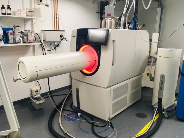
Recruiting an expert for the IVIF agora site
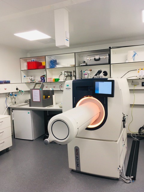
This new 3 Tesla MRI equipped with a cryoprobe and a full set of coils, as well as cutting edge instruments, are waiting for a specialist to take care of them. For job application please follow the CHUV portal.
Happy holiday season
Dear all,
The IVIF team is wishing you a great holiday season. I would like to thank you all for your collaboration and professionalism.
It has been a pleasure to interact and help all of you in your experiments on a daily basis. It has been so diverse and instructive for us.
We will come back with a newsletter in January to show you all the implementations we have made and advancement in in vivo imaging in the institution.
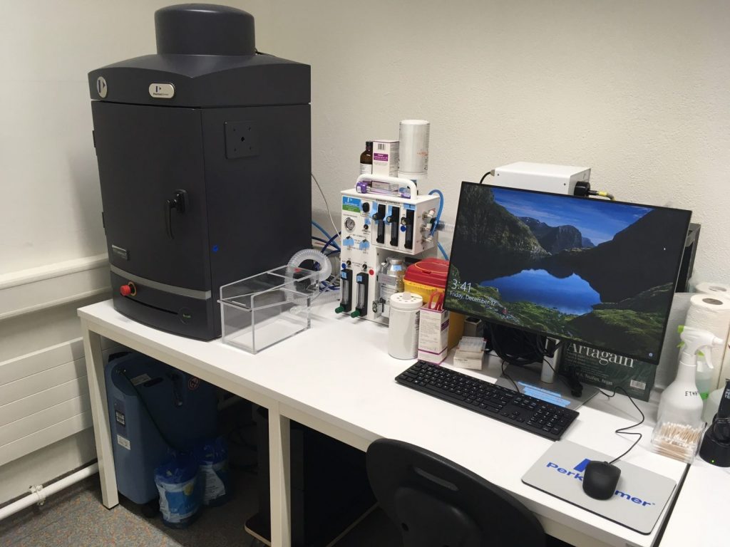
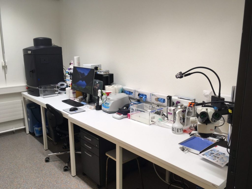
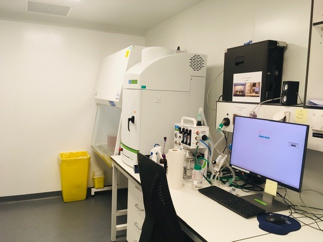
Installation of a new IVIS Lumina III for the CLE
Thanks to an investment from the faculty, I am pleased to announce the installation of a new IVIS lumina III at epalinges, building CLE C room 104A. It will offer a back up for the actual IVIS lumina II.