
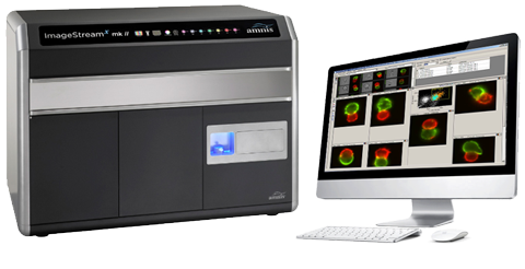
The Amnis ImageStreamX Mark II instrument from Merck Millipore combines high resolution microscopy with flow cytometry. Images of each cell are acquired while they run with high speed (up to 5000 cells per second) through the machine. In addition to obtaining classical flow cytometry data the images dramatically increase the information gained about the cells. For example cell shape, nuclear shape, subcellular localization of proteins and many more features can be analyzed. Most dyes used in flow cytometry can be detected and compensation is applied to the images as well, thus allowing the use of many more colors compared to microscopy. In our 4-laser (405nm, 488nm, 561nm and 640nm) and 2-camera configuration 10 different fluorescent channels can be analyzed simultaneously. In addition three different objectives can be chosen (20x, 40x and 60x) allowing the adaptation of the objective for the respective application. A 96-well plate loader is included allowing the analysis of many samples without being physically present at the machine. Data analysis is performed using the integrated IDEAS software, which allows the analysis of more than 85 parameters per cell. Together with the short acquisition time this allows the analysis of statistically relevant numbers of images, which is not possible using classical microscopy. Typical applications for this machine include autophagy, internalization of proteins, translocation of proteins into the nucleus, cell signaling, analysis of immune synapses, subcellular localization of the protein of interest, cell cycle analysis and many more. Another huge advantage of the Image Stream is the possibility of visual confirmation, for example to determine whether rare events are really cells or not.
In order to use this machine, please contact Kevin Blackney or Francisco Sala de Oyanguren.