PhD projects available
We currently have several PhD projects on cardiovascular MR imaging available. Please find the flyers below:
- Low-Field MRI with Prof. Matthias Stuber:
Are you passionate about advancing healthcare through cutting-edge research? We are offering an exciting opportunity for a highly motivated individual to apply their physics or engineering expertise to a groundbreaking project with broad international impact in the field of medical imaging. Join our dynamic and diverse international team of engineers, mathematicians, and physicists, working closely with medical professionals at the University Hospital in Lausanne.
Master Thesis Projects
Master’s project in fetal cardiac MRI
Background: Advances in fetal cardiac imaging, have contributed to improved decision making and a decrease in the morbidity and mortality of children with congenital heart disease. As a result, there is growing interest in using magnetic resonance imaging (MRI) during pregnancy as a safe diagnostic tool that can compliment the current gold standard ultrasound. The convergence of emerging 3D MRI technology with the unmet challenges of fetal imaging have inspired our group to create a new approach to fetal cardiac MRI. Through the development of fetal-specific dynamic 3D acquisition and reconstruction strategies we aim to create an easy-to-use comprehensive and quantitative diagnostic tool for evaluating the fetal heart in utero.
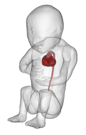
Project Description: This work will combine cutting-edge physics and engineering research with an impactful clinical application. Candidates will be able to familiarize themselves with both the technical (i.e. MR acquisition and reconstruction) and clinical (fetal cardiology) aspects of the project, as we explore the development and optimization of 3D MRI sequences and motion-robust image reconstruction algorithms tailored to imaging the fetal heart. Initial development and simulation will be performed in MATLAB followed by optimization in phantoms and healthy adult volunteers, before scanning in pregnant volunteers.
Location: This Master’s project will take place in the Department of Radiology at the Lausanne University Hospital (CHUV) and the University of Lausanne (UNIL) in Switzerland under the supervision of Dr. Christopher Roy as part of the research team of Prof. Matthias Stuber. You will be part of a group of ~15 engineers and physicists working within the hospital in close collaboration with our clinical partners in Radiology and Cardiology. Our group has access to 4 state-of-the-art clinical MRI scanners, and you will actively collaborate with Siemens Healthcare.
Qualifications: We are looking for highly motivated candidates with a background in engineering, mathematics, physics, life science, or similar. The ability to think creatively and critically within a team environment are required. Computer programming skills are desirable.
To Apply: For further information or to apply (including a CV and motivation letter), please contact Dr. Christopher Roy (christopher.roy@chuv.ch). Project lengths can be tailored to the students’ requirements.
Precision measurements in cardiac MRI
Are you a student in Micro- or Electrical Engineering who wants to complete a Master’s Thesis at the intercept of engineering, medicine, and cutting-edge magnetic resonance imaging (MRI) research?
Advanced, next generation magnetic resonance imaging technology is currently being developed by a team of almost 20 basic scientists and engineers located directly at the CHUV (University Hospital Lausanne). We are working on new technology that visualizes the heart, its structure, its blood vessels and its function with unprecedented detail. In that pursuit, we develop techniques to suppress adverse effects of respiratory and cardiac motion. However, these techniques remain to be characterized and validated not only to better understand – but also to maximize their performance. To meet that challenge, we have already built an MRI-compatible moving phantom whose motion can be programmed. However, the challenge is now to measure the “true” displacement of the phantom head as a function of time with sub-millimeter accuracy. We anticipate that the candidate will implement an optical measurement system that is powered by a laser and which will log phantom motion data during the MRI measurements. Skills in the domains of fiber optics, programming, signal processing, image processing and even experimental scanning on the MRI console will be acquired or further developed during the course of this interesting Masters thesis.
Supervision: Matthias Stuber
Improving quantitative 3D magnetic resonance imaging for the mapping of cartilage damage in knee osteoarthritis
Topic: Magnetic resonance imaging (MRI) numerical simulations, data acquisition, and image processing.
Who: Students with a background in physics or engineering who are interested in a project that will lead to a Master of Science (MSc) degree. Programming knowledge is essential, Matlab knowledge is an advantage.
Where: The project will take place in the Center for Biomedical Imaging at the Lausanne University Hospital (CHUV) and University of Lausanne (UNIL) in Switzerland (see www.unil.ch/cvmr). One of the main goals of the group is to develop new MRI techniques and to apply these in patients in a clinical setting.
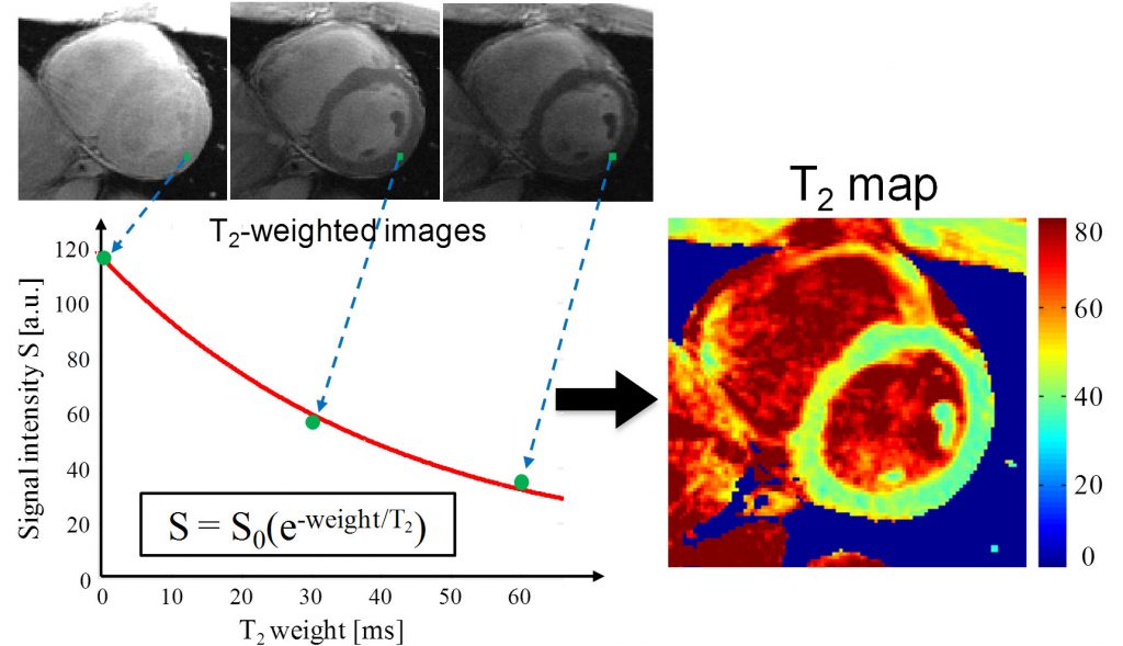
Project: MRI is a non-invasive and non-harmful medical imaging technique. The contrast in its images relies on the difference in so-called relaxation times between tissues. In the context of knee cartilage, the T2 relaxation time correlates with the water and collagen content of the extracellular matrix, as well as the structure of the collagen network. The T2 relaxation time can thus be used as imaging biomarker of early-stage osteoarthritis (OA), during which the breakdown of the cartilage matrix and cartilage loss start occurring. In T2 mapping, the acquisition of multiple images with different T2 weights enables the calculation of the T2 value for each pixel. Unlike the qualitative evaluation of (arbitrary) grayscale intensity images, T2 mapping can potentially help in more accurately, precisely and quantitatively identifying early-stage OA cartilage changes.
Our group recently developed a new 3D technique for knee cartilage T2 mapping with a very high spatial resolution (Colotti et al., J Magn Reson 2017). The Master thesis project will aim to improve this technique in several ways, among others by refining and accelerating the modeling and calculation of the T2 relaxation time. In practice, this means that the student will: 1) familiarize her- or himself with MRI theory and the MRI scanner; 2) expand and improve the numerical simulations of the technique; 3) develop a dictionary T2 fitting framework in Matlab. This improved fitting framework will be then compared with the method that is currently used in terms of precision/accuracy of T2 quantification in phantoms and volunteers. A collaboration with the clinical muskoskeletal radiology service is also foreseen.
Duration: The project duration can be adapted to the student’s requirements, but is expected to take 5 to 9 months.
Supervision: Dr. Ruud van Heeswijk
Accelerated 3D cardiac T2 mapping
Who: We are looking for a highly motivated physics or engineering Master student for a project that will lead to a Master of Science (MSc) degree.
Where: The project will be on the subject of magnetic resonance imaging (MRI), and will take place in the CardioVascular Magnetic Resonance center (CVMR, see www.unil.ch/cvmr) of the University Hospital of Lausanne (CHUV) and the University of Lausanne (UNIL) in Switzerland.
What: The goal of the project will be to expand a two-dimensional (2D) T2 mapping technique to 3D.
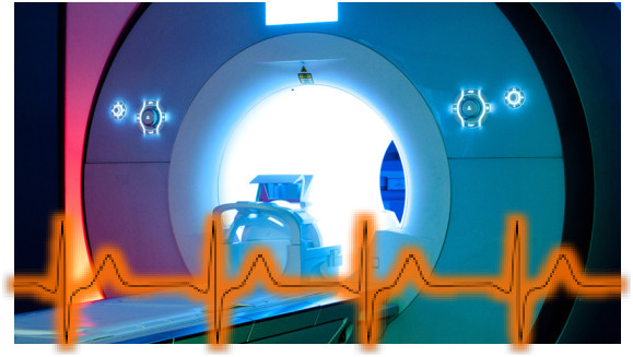
Background: Magnetic resonance imaging (MRI) is a non-invasive medical imaging technique without ionizing radiation. MRI contrast between tissues is mostly determined by the T1 and T2 relaxation times, which depend on the composition of the tissue that is being imaged (water, fat, muscle, etc.). The T2 relaxation time has the unique property that it changes if the tissue becomes swollen (edema), which is the case in myocardial infarction for instance. In so-called T2 mapping, multiple images with different T2 weights are used to calculate the T2 value for each pixel. This quantitative technique with values instead of arbitrary grayscale intensities can therefore potentially help in more accurately and precisely identifying edema and thus to distinguish treatable heart muscle from dead tissue.
The CVMR recently developed a technique that combines T2 mapping with a KWIC filter that shares part of the data between different T2-weighted input images in order to accelerate the acquisition time. This new technique called SKRATCH was developed as a 2D acquisition (i.e. a single slice). The objective of this project is thus to convert the existing technique to 3D acquisition.
The work itself: The project will consist of first familiarizing oneself with MRI theory and the MRI scanner, establishing and optimizing the combination of the different T2-weighted images via Matlab, defining a 3D T2 mapping protocol in vitro (bottles of water with predetermined T2 values) that is processed in MATLAB, followed by MRI scans in healthy volunteers. Collaboration with the clinical cardiology service is also foreseen.
Duration: The project duration can be adapted to the student’s requirements.
Supervision: Dr. Ruud van Heeswijk