This novel Swiss National Science Foundation (SNSF)-funded (SNSF #320030_173129) technology (Di Sopra et al. MRM 2019)1 1 deliberately removes contemporary hurdles in CMR including operator dependency and time inefficiency (many contemporary methods sample data during 2 percent of the time only). The Free-Running Framework obviates the need for ECG triggering, ECG lead placement, breath-holds, and navigators. This shifts the paradigm from a prospective scan parameter- and imaging plane selection to a single-click solution where 3D data can freely be queried for structural and functional information retrospectively and after the scan. This significantly reduces operator dependency. As for time efficiency, data are no longer collected at regular intervals synchronized to the heartbeat, but they are rather collected continuously with high temporal resolution and irrespective of the heart’s position or contractile state. Motion consistent data throughout the cardiac cycle and for multiple respiratory levels are then simply extracted after the fact. Arguably, no other CMR technology collects more high-resolution 3D data of the heart per unit time, and the scan efficiency has improved more than 15-fold.
History
The development of this framework is tightly linked with the advent of “self-gating”, where respiratory and cardiac signals are directly extracted from the acquired k-space data used for imaging, rather than from external devices such as the ECG, respiratory sensors, or navigators. Landmark publications include those by Kim et al. (MRM 1990)2, Hardy et al. (MRM 2003)3, Larson et al. (MRM 2004)4, Stehning et al. (MRM 2005)5, Pang et al. (MRM 2014)6, and Coppo et al. (MRM 2015)7. With the advent of compressed sensing (Lustig et al. MRM 2007)8 and motion-resolved reconstructions by Feng et al. (MRM 2016)9, the way was paved for respiratory-resolved 4D reconstructions (Piccini et al. MRM 2017)10 and by extension for respiratory- and cardiac motion-resolved 5D reconstructions (Feng et a. MRM 2018)11. By combining the free-running bSSFP sequence of Coppo et al. (MRM 2015)7 with a sophisticated cardiac- and respiratory self-gating signal extraction to inform binning of a k-t-sparse SENSE image reconstruction, the “Free-Running Framework” was born (Di Sopra et al. MRM 2019)1 and has since been extended with FISS (Bastiaansen et al. MRM 2020)12 and LIBRE (Masala et al. MRM 2020 In Press) to provide more effective fat suppression, and with flow encoding capabilities to enable simple and time-efficient 5D flow (Ma et al. ISMRM 2019) measurements.
Technology
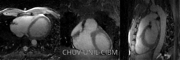
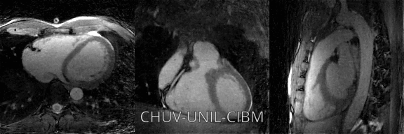
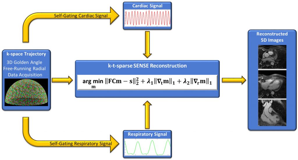
 The Free-Running Framework rests on three major pillars. The first pillar includes a non-interrupted free-running sequence that continuously collects 3D data of the heart during a few minutes regardless of the heart’s contractile state or the respiratory position. A 3D radial trajectory that is continuously being rotated by the golden angle forms one of the central ingredients. This sequence has been implemented at 1.5T and 3T as either a bSSFP or a gradient echo version. Contrast agents are an option but not a requirement. The spatial resolution of this method currently ranges from 0.9-2.5mm, the sampling efficiency is 30 percent, and fat signal saturation can either be obtained with conventional fat suppression (Coppo MRM 2014) 7, FISS (Bastiaansen MRM 2020) 12, or LIBRE (Bastiaansen MRM 2018 13, Bastiaansen MRM 2019 14, Masala MRM In Press 2020). Sophisticated signal processing forms the second pillar and enables physiological signal extraction from the acquired data rather than from external gating and triggering devices (Di Sopra et al MRM 2019) 1. With these physiology signals, motion-consistent sub-sets of the free-running data are extracted and assigned to respiratory and cardiac bins. By exploiting sparsity in the cardiac and respiratory dimensions among these bins, compressed sensing k-t-SPARSE SENSE reconstruction, our third pillar, leads to more than 100 cardiac- and respiratory motion-resolved 3D images per dataset.
Applications
At this juncture, the free-running framework supports anatomical and functional imaging of the whole heart with high temporal and spatial resolution. However, and owing to the modular structure of this concept, our team has now begun to deliberately expand the free-running framework to 5D flow imaging (Collaboration Dr. Michael Markl and Dr. Liliana Ma Northwestern University Chicago, Chicago, IL, USA). In parallel, new avenues for 5D tissue mapping and fat fraction mapping are independently being explored by the teams of PD Dr. van Heeswijk and Dr. Bastiaansen. Together with Dr. Mark Fogel and Dr. Kevin Whitehead from the Childrens Hospital of Philadelphia, Philadelphia, PA, USA, our framework enables highly detailed anatomical and functional imaging of the heart in pediatric patients. At the same time, high-resolution structural imaging of the heart to improve our understanding of arrhythmias is pursued by Dr. Jerome Yerly in collaboration with Dr. Hubert Cochet, Dr. Pierre Jaïs, Dr. Bruno Quesson, and Dr. Aurélien Bustin from LIRYC, University of Bordeaux, France.
Awards & Distinctions
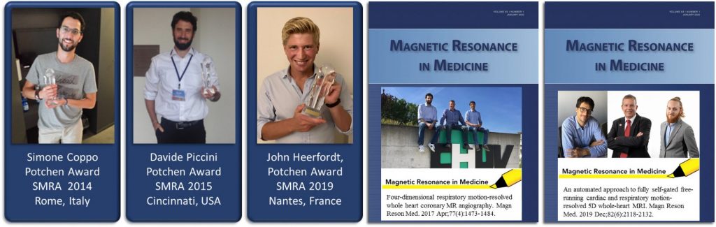
In 2014, Simone Coppo was bestowed the Potchen Award for his free-running sequence at the annual meeting of the SMRA in Rome, Italy. In the following year in Cincinnati, Davide Piccini was the winner in the same category for his work on motion-resolved reconstruction, which was also selected for the MRM Editor’s Pick in 201715. In 2019, at the SMRA in Nantes, John Heerfordt was handed the Potchen Award for his SIMBA technology that builds on the free-running framework. In 2019, the MRM paper of Lorenzo di Sopra and Jerome Yerly that describes the free-running framework1 was the MRM Editor’s Pick in December.
Literature
1. Di Sopra L, Piccini D, Coppo S, Stuber M, Yerly J. An automated approach to fully self-gated free-running cardiac and respiratory motion-resolved 5D whole-heart MRI. Magn Reson Med. 2019;82(6):2118-2132.
2. Kim WS, Mun CW, Kim DJ, Cho ZH. Extraction of cardiac and respiratory motion cycles by use of projection data and its applications to NMR imaging. Magn Reson Med. 1990;13(1):25-37.
3. Hardy CJ, Zhao L, Zong X, Saranathan M, Yucel EK. Coronary MR angiography: respiratory motion correction with BACSPIN. J Magn Reson Imaging. 2003;17(2):170-176.
4. Larson AC, White RD, Laub G, McVeigh ER, Li D, Simonetti OP. Self-gated cardiac cine MRI. Magn Reson Med. 2004;51(1):93-102.
5. Stehning C, Bornert P, Nehrke K, Eggers H, Stuber M. Free-breathing whole-heart MRI with 3D-radial SSFP and self-navigated image reconstruction. Magn Reson Med. 2005;In Press.
6. Pang J, Bhat H, Sharif B, et al. Whole-heart coronary MRA with 100 percent respiratory gating efficiency: self-navigated three-dimensional retrospective image-based motion correction (TRIM). Magn Reson Med. 2014;71(1):67-74.
7. Coppo S, Piccini D, Bonanno G, et al. Free-running 4D whole-heart self-navigated golden angle MRI: Initial results. Magn Reson Med. 2015;74(5):1306-1316.
8. Lustig M, Donoho D, Pauly JM. Sparse MRI: The application of compressed sensing for rapid MR imaging. Magn Reson Med. 2007;58(6):1182-1195.
9. Feng L, Axel L, Chandarana H, Block KT, Sodickson DK, Otazo R. XD-GRASP: Golden-angle radial MRI with reconstruction of extra motion-state dimensions using compressed sensing. Magn Reson Med. 2016;75(2):775-788.
10. Piccini D, Feng L, Bonanno G, et al. Four-dimensional respiratory motion-resolved whole heart coronary MR angiography. Magn Reson Med. 2016.
11. Feng L, Coppo S, Piccini D, et al. 5D whole-heart sparse MRI. Magn Reson Med. 2018;79(2):826-838.
12. Bastiaansen JAM, Piccini D, Di Sopra L, et al. Natively fat-suppressed 5D whole-heart MRI with a radial free-running fast-interrupted steady-state (FISS) sequence at 1.5T and 3T. Magn Reson Med. 2020;83(1):45-55.
13. Bastiaansen JAM, Stuber M. Flexible water excitation for fat-free MRI at 3T using lipid insensitive binomial off-resonant RF excitation (LIBRE) pulses. Magn Reson Med. 2018;79(6):3007-3017.
14. Bastiaansen JAM, van Heeswijk RB, Stuber M, Piccini D. Noncontrast free-breathing respiratory self-navigated coronary artery cardiovascular magnetic resonance angiography at 3 T using lipid insensitive binomial off-resonant excitation (LIBRE). J Cardiovasc Magn Reson. 2019;21(1):38.
15. Piccini D, Feng L, Bonanno G, et al. Four-dimensional respiratory motion-resolved whole heart coronary MR angiography. Magn Reson Med. 2017;77(4):1473-1484.