|
|

|
|
|
|
|
|
|
Facility News
Oh yeah, September is here and I am fairly certain it brings back a lot of first day of school 🏫 memories where waking up was nearly impossible after so many weeks of holidays ! Aren't you glad you don't get to have vacation and can keep enjoying the thrill of Science 🔬 all year round ?? As your FCF Staff, we're certainly happy to be here and support you for it !
|
|
Deborah Lecuyer won the mug of the month below, congratulations to you ! Please come by to our office at the SE-C Biopole to pick it up !
|
|

|
Please try to answer our quiz it and get a chance to win our next mug of the month ! Good luck to you all !
|
|
|
|
|
|
FACS Tips
|
Who Killed the Double-Positive? A Flow Cytometry Whodunit
|
The Case
Back in August of 2010, a paper was published in the journal Blood that had come to an exciting new discovery.
|
|
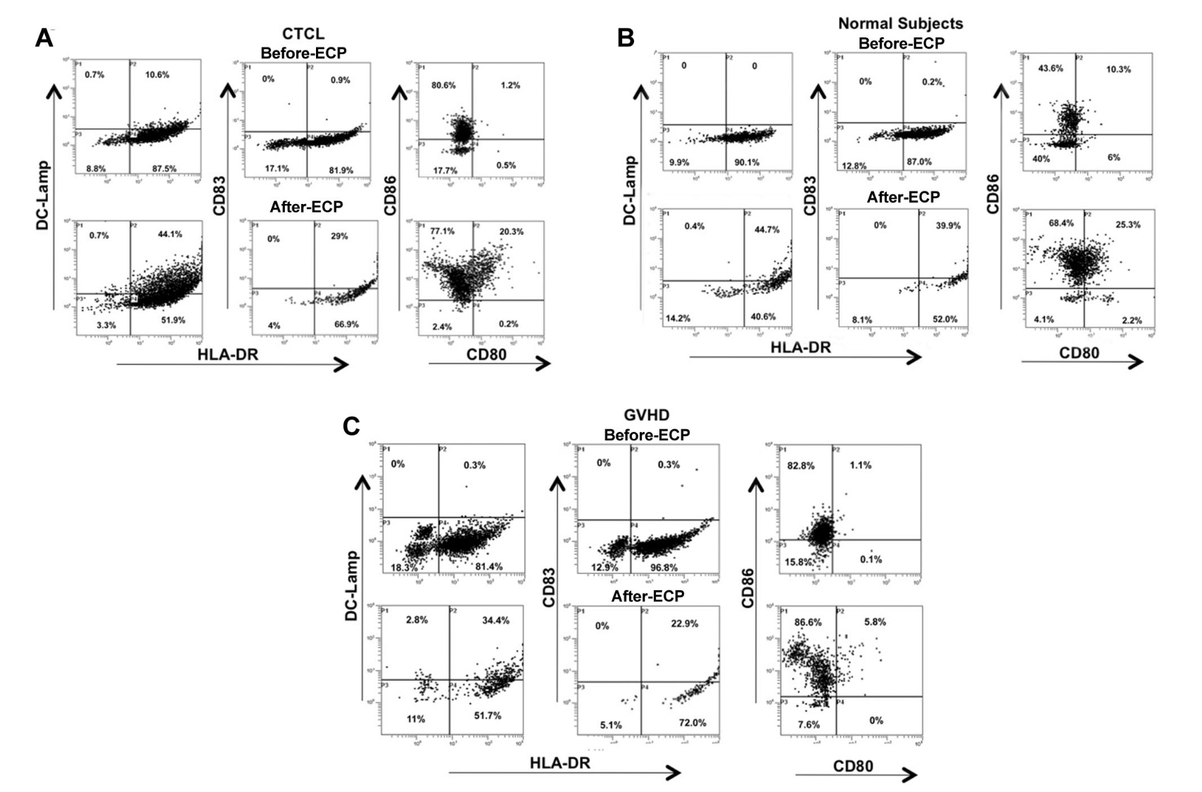
This discovery was backed by flow cytometry showing the monocyte-to-DC transition before and after ECP treatment across normal and disease-state subjects as demonstrated below.
|
|
|

|
Upper-right quadrants highlight the double-positive population (HLA-DR+CD83/DC-LAMP+) indicating newly matured DCs. Pre-ECP samples show minimal double-positive cells; post-ECP (Day 1) samples show prominent populations. The tight clustering of post-ECP events suggests maturationally synchronized DCs, distinct from the heterogeneous scatter typical of 6-day conventional cultures with GM-CSF and IL-4. All three groups, regardless of disease state, showed similar induction.
|
The Challenge
But not everyone was convinced, and one skeptic carried lofty credentials. In January 2011, Mario Roederer and James F. George published a letter in Blood raising a pointed technical question: could the entire result be an artifact of improper compensation?
|
|
Roederer is no ordinary reviewer. He authored one of the foundational paper on compensation theory and is widely recognized as one of the world's leading experts in multicolor flow cytometry.
|
|
|
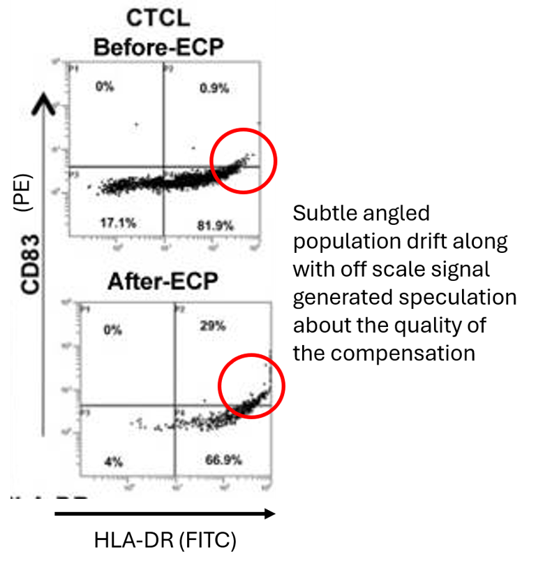
In the letter, James F. George and Mario Roederer, argued that this double positive population increase could have been the result of improper compensation. Their explanation was that, when one marker (HLA-DR) is expressed at very high intensity, so high that it becomes off-scale after ECP treatment, and another (CD83) at lower intensity are in the same sample, improper fluorescence compensation can artificially inflate the apparent positivity of the lower-intensity marker.
|
|
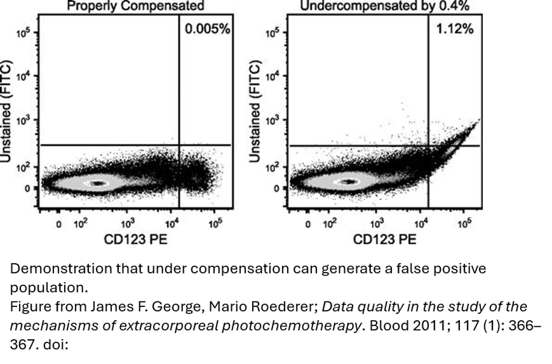
The problem with off scale data, is it is not possible to properly compensate it, and in addition to that, rules of compensation state that a single stain should be as bright or brighter than the fully stained sample, which would be problematic here, if the data were already off scale. The likely result of this would be an under compensation effect leading to a false positive staining into the CD83 channel. This is backed up, they argue, by the narrow distribution skewing upward at a 45° angle at high HLA-DR expression levels.
|
|
|
In the example provided by George and Roderer above, they show how improperly compensated data could create a false positive population in the unstained FITC channel from PE, these are the same fluorophores used in the published experiment by Berger et al.
|
They also think it’s possible there may be an overcompensation error in the original paper between CD80 and CD86.
|
Finally, they argue in their letter that gates should have been set off FMO controls, especially when dealing with co-expressing markers, rather than using quadrants.
|
The Defense
In a response again published in Blood, the original authors stood behind their work, and claim the data was compensated correctly. They also add that they had done parallel tests with similar results in single colour experiments, negating this possible error type. Also, they had further molecular and functional studies beyond their flow cytometry results to confirm their original hypothesis.
|
The Conclusion
At this point, there were no further commentaries on this project, and has no retraction or correction notice. But here's what's important: George and Roederer were right to be skeptical. The flow data presentation did have real problems, off-scale values, compensation questions, gating choices that deserved better documentation. These were genuine weaknesses that probably should have been fixed or at least acknowledged in the original paper.
|
|
Fortunately for Berger and colleagues, their solid data elsewhere backed up everything they had claimed. However, it is a good message to ensure the fundamentals in your flow cytometry experiments are sound. Little issues like improperly scaled gains/voltages, or compensation errors could have illegitimized the entire results of this study if that was the sole footing of this project. If you have questions about any of your flow cytometry results, as always feel free to reach out to the staff at the FCF.
|
|
|
|
|