|
|

|
|
|
|
|
Facility News
This month we have a big news for the Agora ! By the end of the month, the Cytek Aurora 3 will be officially available ! Dear Agorians, be ready to explore spectral cytometry 🌈 !
|
|
As a reminder,We will have a day long workshop on October 15th about imaging flow cytometry and cell sorting (DETAILS HERE). As some of you may know, we acquired a BD Discover S8 spectral sorter recently and it offers new options for your experiments. Please join us for a morning presentation followed by an hands-on session in the afternoon ! Contact us if you'd like to submit some samples !
|
|
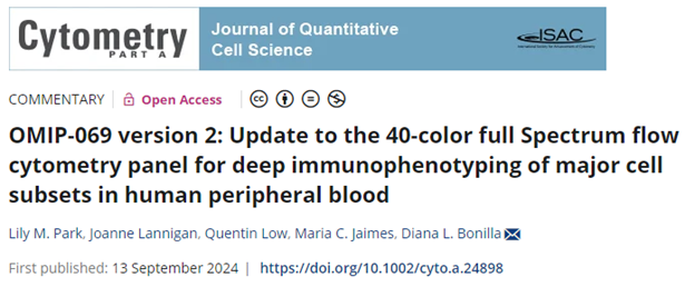
In this month FACS Tips, Kevin is going back to OMIPs since one of them has been updated for the first time ever ! A great read on how to always strive to improve your flow !
|
|
Romain Veber won the mug this month, Congratulations to you !
|
|
Each month, we will ask you 3 questions about the newsletter topic. If you win, you can enter the lottery to win a unique mug designed by the FCF team !
Please take few minutes to answer the quiz HERE.
|
|
|
|
|
FACS Tips
|
Version 2: Remixing the Benchmark
|
When we think of gold standards in panel design, OMIPs often come to mind. These fully optimized panels are meticulously developed, with every antibody titrated and every instance of spillover accounted for. While I’ve previously covered OMIPs (Optimized Multicolor Immunofluorescence Panels) in more detail, as a brief reminder: OMIPs are a collection of publications (first appearing in Cytometry Part A in 2010) that provide researchers with templates for reproducing complex staining panels. They significantly reduce troubleshooting and setup time, making them incredibly valuable research tools. For those building large or tissue-specific panels, OMIPs can save both time and money by offering a pre-established framework. However, as with most things, it’s not always that simple.
|
“But Mouse, you are not alone, in proving foresight may be vain: The best-laid plans of mice and men often go awry”.
|
|
Though written nearly 200 years before the invention of flow cytometry, Robert Burns seems to have perfectly captured the transition many of us face when moving from design to data in our experiments.
|
|
|
At the time of its publication in 2020, OMIP-069 was a big step forward in spectral cytometry. The first OMIP to surpass 30 colours, this 40 colour Deep Immunophenotyping panel designed for Human peripheral blood samples was highly cited and since has been adopted in full or part by various laboratories. Yet, through its widespread adoption, the limitations of the panel began to surface. So, the publishers returned to the lab, and this month have published the first ever OMIP update, OMIP-069 Version 2. This update targets two key issues: reagent availability and performance.
|
|
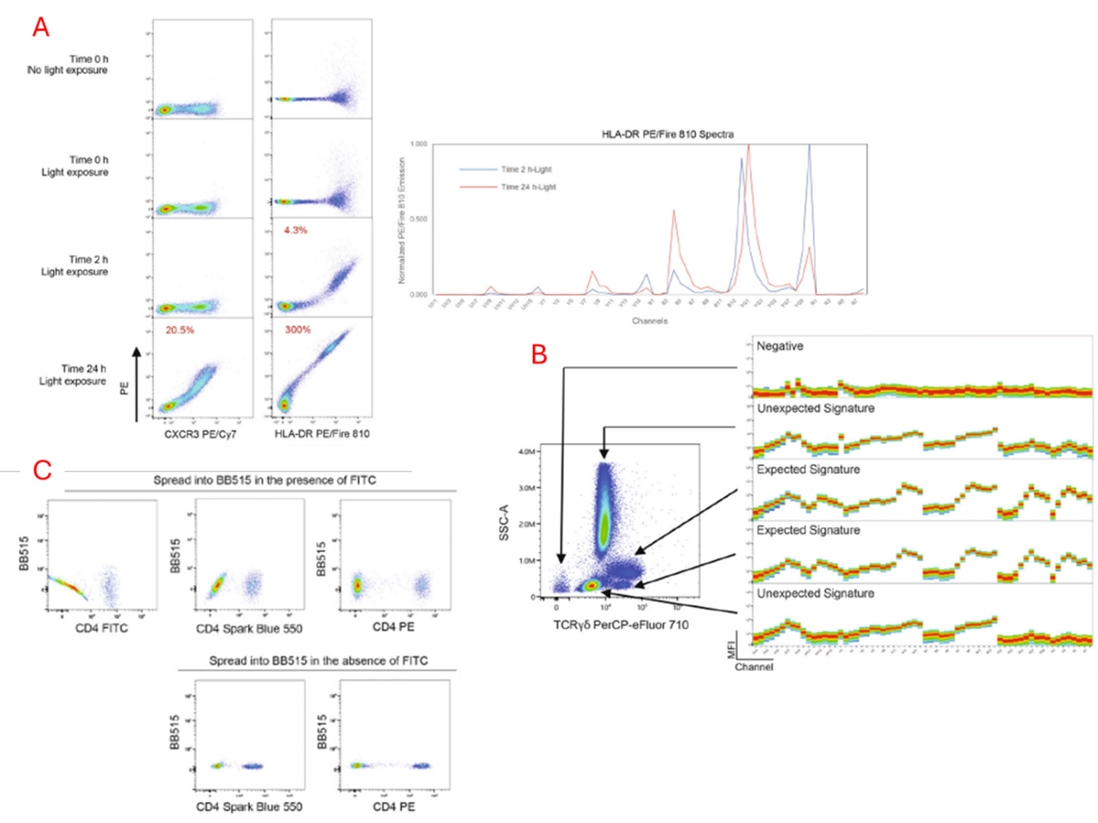
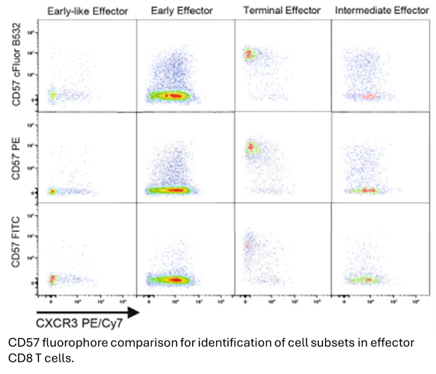
Firstly, some antibodies included in the original panel were consistently hard to purchase, languishing in back orders for too long to be useful. Next, certain antibodies showed performance issues. One notable example mentioned in the paper was HLA-DR PE/Fire 810, which exhibited stability problems (A). Further experimentation revealed that this marker was more sensitive to degradation than typical tandem dyes, especially after two hours of light exposure, which caused increased emission peaks in the base PE fluorophore. While this fluorophore could be used with more caution it was determined it was better to replace it. Another challenging fluorophore from the original panel was TCRγδ in PerCP-eFluor 710, which displayed multiple spectra in an inconsistent manner across lots (B). This had a profound effect on unmixing in various situations. Lastly, the use of FITC and BB515, which have heavy spectral overlap and a similarity score of 0.98, were not as reliably unmixed in replication by others as shown in the original paper (C). To address these two branches of problems, the publishers rigorously vetted alternatives to overcome these issues with minimal alteration to the original panel and gating strategy.
|
|
|
|
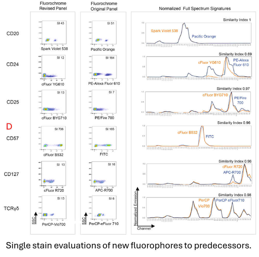
In total, eight markers were moved to alternate fluorophores in the updated panel. Six of these were simply moved to near identical emission spectra fluorophores to maintain consistency (D). For example, cFluor 532 was tested as a replacement for FITC to address the high spillover between BB515 and FITC. While FITC and cFluor 532 have a similarity index of 0.96, the similarity between BB515 and cFluor 532 is only 0.89, down from 0.98 with FITC. This adjustment fixed the significant spreading error. The remaining two had more spectrally dramatic alterations with HLA-DR PE/Fire 810 moving to BV480 and IgD BV480 moving to cFluor BYG750. Each new fluorophore was compared to its predecessor for staining patterns and resolution, using optimally titrated single stains.
|
|
|
|
Once it was determined each marker produced similar or improved results as a single stain, a further assessment of a smaller panel to distinguish its ability to reliably and similarly resolve subsets was performed. This was done for both T cell and B cell subset markers as an optimal titer. Interestingly, CD24 in eFluor YG610, although it performed poorer on single stain evaluation for stain index compared to predecessor, had greater spatial staining resolution on the small panel test and it appears the new marker had less non-specific binding.
|
|
|
|
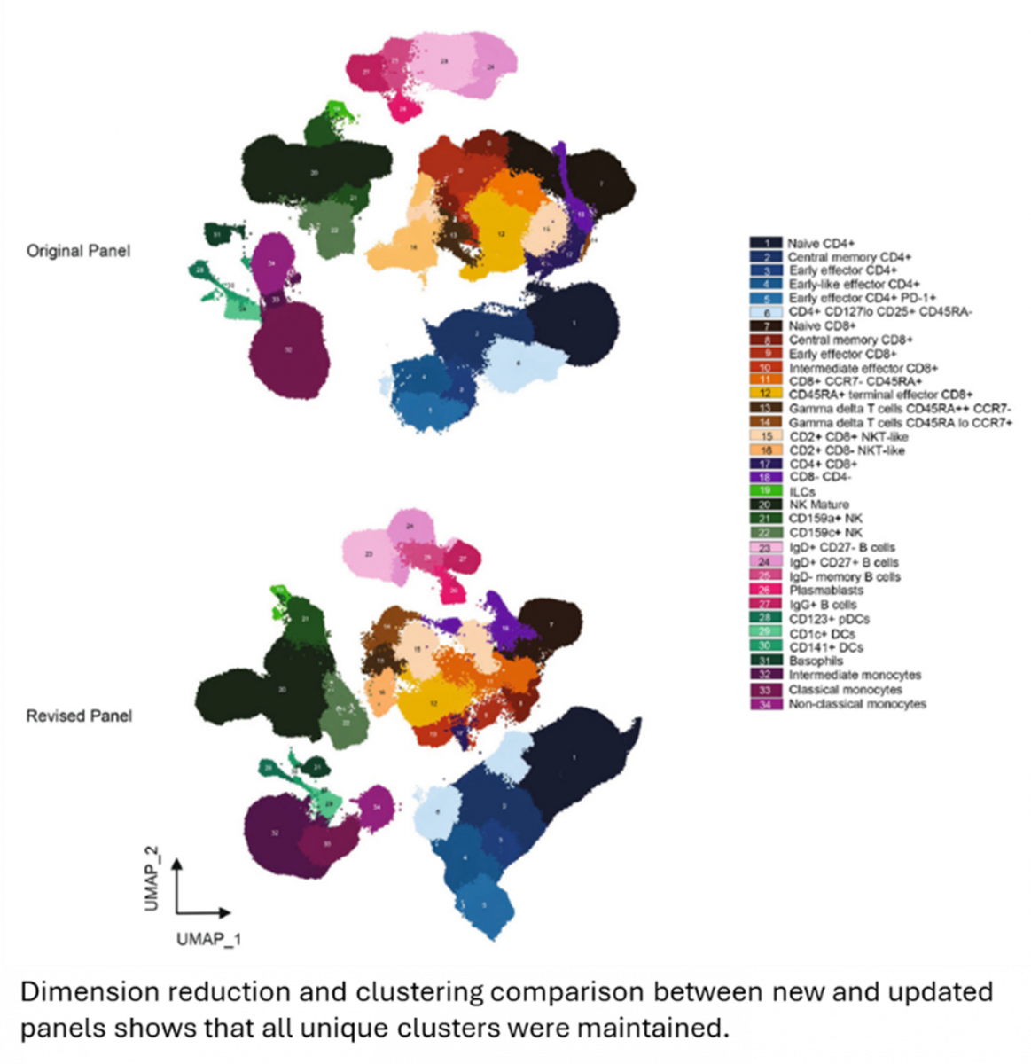
Finally, a fully stained test was performed. Using manual gating to identify each population, and then performing a dimension reduction and clustering analysis, all the previously identified populations could be properly resolved again as unique clusters with the updated panel.
|
|
|
This serves as a reminder that even when we’ve accounted for everything in panel design, unforeseen challenges can arise, requiring adaptation. While this article may cover a more complex case than our usual day-to-day panel adjustments, it offers a valuable framework for making changes. The authors also leave open the possibility that similar updates may occur for other OMIPs in the future, reflecting new advancements or addressing issues. The key takeaway: just because a panel is well-established doesn't mean it can't be improved. With careful attention to marker performance and fluorophore compatibility, panels can be refined without undermining the work that came before.
|
|
As always, feel free to reach out to the FCF if you have any questions about your flow cytometry experiments.
|
|
|
|
|