|
|

|
|
|
|
|
|
|
Facility News
Our facility is welcoming new members ! The Cytoflex LX5 & LX6 are our newest recruits and will surely help our users to find a machine anytime they need one !
|
|
They have a 5 laser configuration that can be found on our website LX5 & LX6. You can easily run 10-12 colors panel in 96 well plates automatically while you can focus on other tasks during your day ! So give them a try if you've never tried and for the current Cytoflex lovers, the machines are already accessible to you !
|
|
|
This month, Kevin is presenting useful techniques and reagents to reduce non specific signals in your staining and increase the quality of your data.
|
|
Victor Joo won the mug this month, Congratulations to you !
|
Each month, we will ask you 3 questions about the newsletter topic. If you win, you can enter the lottery to win a unique mug designed by the FCF team !
Please take few minutes to answer the quiz HERE.
|
|

|
|
|
FACS Tips
|
Block and Preserve: Optimized Guide to Preventing Non-Specific Signal
|
|
|
When we stain for Flow Cytometry, we expect that what’s positive is what we targeted, and that makes us certain in our data. To achieve this, we need to take steps to prevent our antibodies from binding non-specifically or losing the expected signal that could affect our results. This can be a difficult task to get right, especially with ever growing panels and new fluorophores with unique and different properties. But when we successfully prevent it, the signal-to-noise ratio improves dramatically, and so does sensitivity.
|
|
A great new paper in Current Protocols by Oliver Burton (who also writes for the fantastic flow cytometry site Colibri Cytometry) tackles this problem head-on (HERE ). Burton and his team developed an optimized strategy to reduce non-specific binding while preserving true signal, with a focus on high-parameter flow cytometry. Even if you work with smaller panels or do intracellular staining, you’ll find their insights highly relevant.
|
|
The Cause
|
|
|
The paper outlines three main contributors to non-specific binding and signal loss:
|
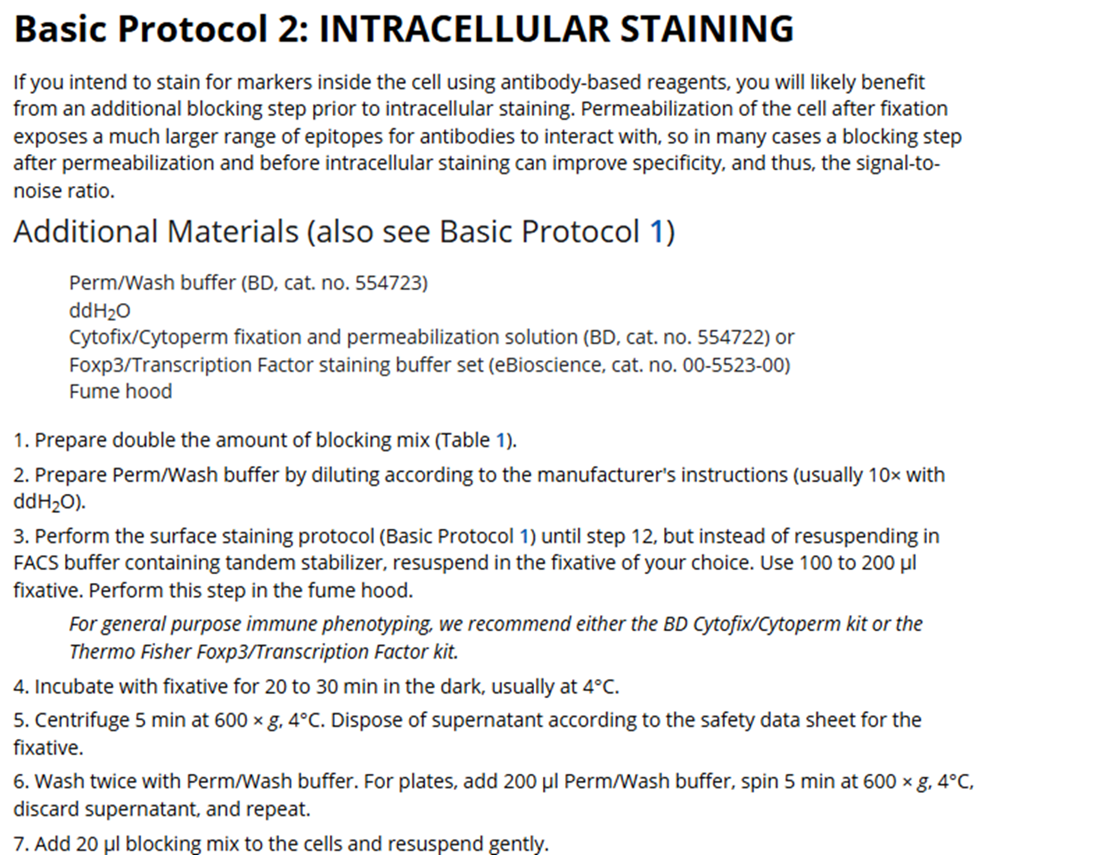
- Fc receptors - Fc receptors naturally bind to the constant (non-variable) region of immunoglobulins. CD16 and CD32 are low-affinity receptors, while CD64 is high-affinity and a major contributor to non-specific interactions. The extent of Fc-mediated binding depends heavily on receptor expression, cell activation state, and the antibody host/isotype used. This type of non-specific binding only occurs on certain cell types, making it unpredictable without proper controls, such as monocyte staining markers.
- Dye interactions - This cell-independent form of non-specific binding occurs mainly with Brilliant dyes, NovaFluors, and Qdots. Their polymer backbones can stick to one another, leading to artificial signal “bridging.” Though less common than Fc-binding, this can cause subtle but serious issues in spectral unmixing and data interpretation.
- Tandem Degradation - Separate from non-specific binding, tandem dye breakdown creates equally problematic artifacts. For example, if the linker between PE and Cy5 breaks, a PE-Cy5-positive cell may appear PE-only, resulting in incorrect marker assignment. This can be a difficult problem to recognize correctly when evaluating your unmixing.
|
The Plan/Protocols
|
|
|
The authors present well outlined protocols for basic surface staining but also detailed instructions for intracellular and cytokine staining, along with some tips and common pitfalls. There is even a guide for making an optimized antibody master mix solution. Here I will highlight some of their main points.
|
|
Fc/Monocte Blocking
|
Including moderate amounts of protein (e.g., BSA 2-5%, FBS 0.5-1%, or FCS 2-5%) in staining buffers helps reduce non-specific antibody binding and background autofluorescence while maintaining cell viability, whereas excessive protein levels can disrupt binding and increase autofluorescence.
|
|
As mentioned, many times before, for many reasons, titrations are an essential part of panel design/experiment preparation. Just by titrating down antibodies, non-specific binding can be reduced. If your panel happens to include an FMO along with staining for monocyte markers, you could even evaluate to what degree background staining can be attributed to non-specific binding from Fc receptor+ monocytes vs say B cells.
|
|
|
In their tests in the paper, the addition of serum (normal serum of the host species for the antibodies being used, or at least of the species that is most used) out performed commercial blocking reagents, especially when staining for human Immunoglobulins.
|
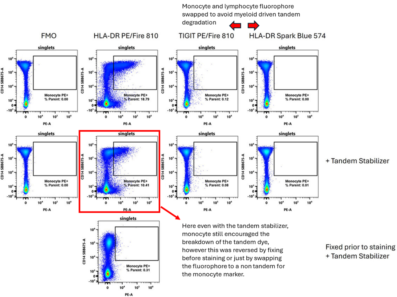
Monocyte blocking reagents (e.g., Monoblock) have become increasingly used as they can prevent non-specific cyanine tandem binding to myeloid cells. However, the authors noted that they can reduce cell recovery. And while they are effective in human samples, murine samples seem far less beneficial, and simply by fixing cells before staining you could prevent entirely this non-specific binding between cyanine tandems and monocytes.
|
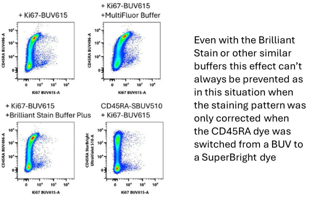
Do not include monocyte blocking reagents in your protocol after perm/fix in your transcription factor staining protocol as the authors saw a substantial loss in target transcription factors after blocking with this reagent post-fix.
|
Dye Interactions
|
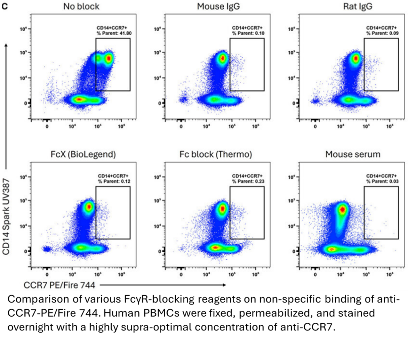
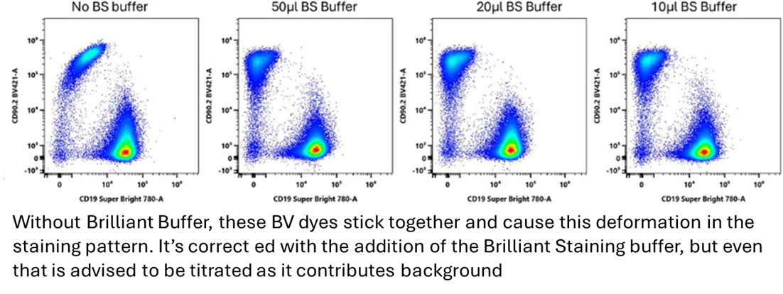
It is well known that the Brilliant dye families, particularly the ultraviolet dyes, have a unique habit of non-specifically binding to one another. While this effect isn’t seen with all conjugates to the same extent, it is something that needs to be considered when using 2 or more of these dyes in a panel.
|
|
|
|
The simplest way to combat these effects is to include a Brilliant Staining buffer. Though, since these buffers do increase the background in channels around BV421, it is advisable to do a titration of the buffer as well. Sometimes swapping to a different, non-interacting fluorophore is required.
|
|
|
Tandem Dye Degradation
|
Tandem breakdown is a result of many things including light, heat, and fixatives. To combat this problem, a tandem stabilizer reagent is now available to add to your buffer as well as your master mix solution.
|
|
|
One other possible solution for fixing this problem is panel design. Focus putting tandems prone to degradation on lymphocytes as opposed to the more metabolically active and degrading myeloid lineage cells. This is likely because monocytes, through their ROS production, catalyze the breakdown of tandems. This problem of tandem degradation can also be solved by fixing the cells first before staining with tandem dyes as that also cuts out the metabolic activity that causes the degradation.
|
Conclusion
If you’ve struggled with unexplained background staining or weak marker resolution, Burton et al.’s work is worth a deep read. With the constant push for larger, more complex panels, it’s refreshing to see a paper that focuses on the fundamentals, improving data quality by addressing non-specific binding and signal preservation.
|
Check out the paper for full protocols and visit Colibri Cytometry for more excellent flow cytometry tips and tricks.
|
|
If you have questions about your staining protocol or need help troubleshooting non-specific staining in your data, reach out to us at the FCF, we’re happy to help!
|
|
|
|
|