|
|

|
|
|
|
|
Facility News
Happy New Year to you all ! I hope 2025 can bring you everything you've dreamed of ! In my case, I dream of matching single color controls, FMOs and experimental controls. I know, boring but very true 😉
|
|
Cheers 🥂 to another year with you all !
|
|
|
|
In this month FACS Tips, we start the year with a different kind of topic. It still revolves around light and colors so you won't get too lost ! Thanks Kevin for exploring new horizons !
|
|
Jan-Pieter Eversdijk won the mug this month, Congratulations to you !
|
|
Each month, we will ask you 3 questions about the newsletter topic. If you win, you can enter the lottery to win a unique mug designed by the FCF team !
Please take few minutes to answer the quiz HERE.
|
|

|
|
|
FACS Tips
|
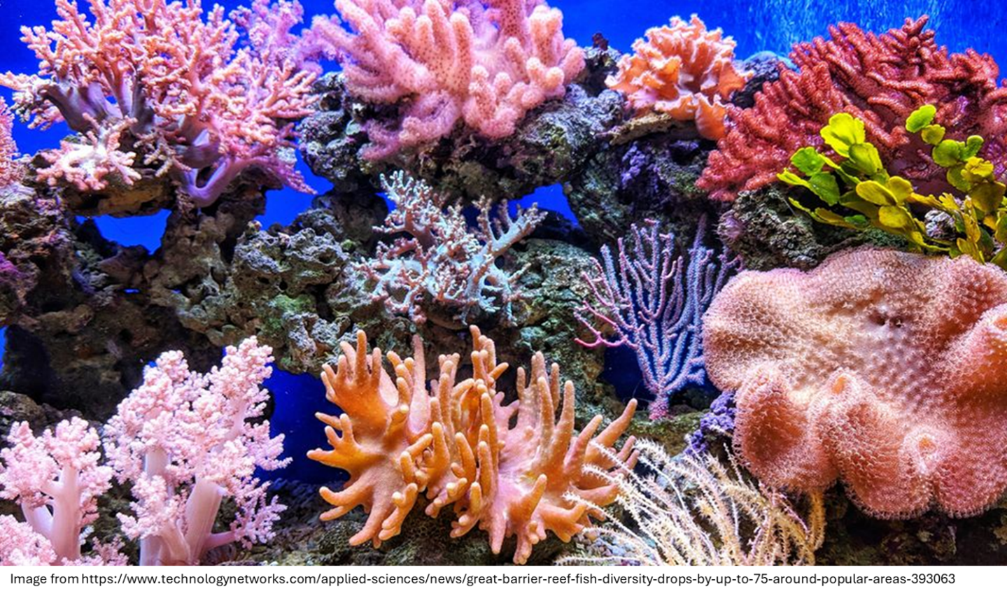
Coral, Nature's Flow Cytometer
|
Flow cytometry can feel hard to explain at times, it's so niche and specialized, requiring expensive synthetic reagents, paired with even more expensive machines, to explore microscopic markers, all through the excitation and emission of lights hidden inside a box. But there is one great example of a natural system that behaves not too differently. Coral. A beautiful rainbow of colours excited under ocean filtered light and fluorescing in unique ways that tell a story about the health and activity of its host. These colorful paradises create an intriguing parallel between what we do in the lab and what happens under the ocean.
|
|
|
When we think of corals, we might picture something like the Great Barrier Reef in Australia—a massively diverse ecosystem home to thousands of species. However, the coral itself is made up of individual animals called polyps that interconnect to form larger colonies. Reef-forming stony coral polyps have a transparent body over a hard, white skeleton. Within these transparent bodies, an essential symbiosis is formed.
|
|
Zooxanthellae, a photosynthetic organism, lives inside the polyp in the millions. It produces the energy that helps the coral grow, while the coral provides a safe environment to shelter this single-celled eukaryote. Together, they create the fluorescent colors we associate with coral reefs.
|
|
|
|
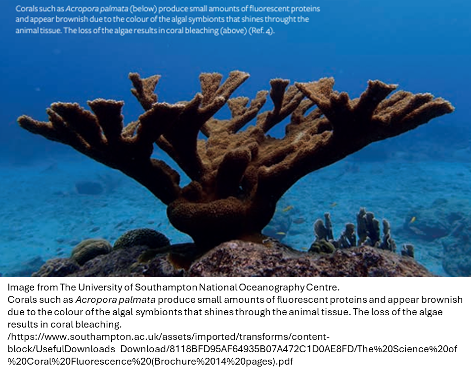
Zooxanthellae, due to its chlorophyll, will fluoresce a yellow-green to brown colour, it’s peak excitation is in the range of 350 to 450nm and it has a peak emission at 683nm. In corals that produce little of its own fluorescent proteins, the zooxanthellae largely determines their colour as in the image above. So, what about all the bright colours?
|
|
|
|
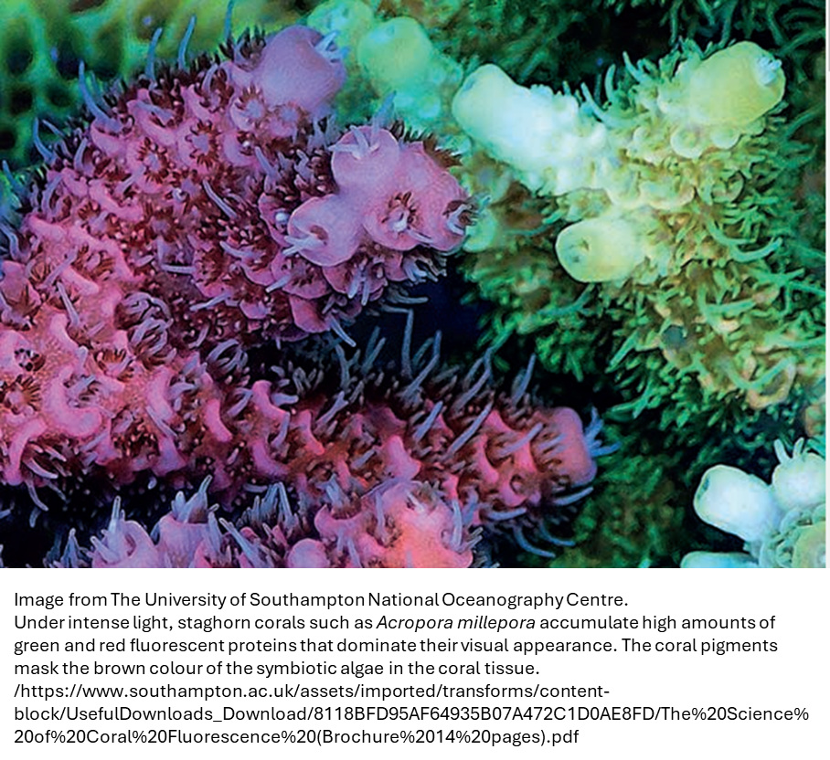
Coral polyps contain special pigments, including more than 85 fluorescent proteins homologous to GFP. Interestingly, the GFP we use in labs was synthesized from the jellyfish Aequorea victoria, not coral. These fluorescent proteins excite and emit at various wavelengths, producing colors such as cyan, green, yellow, and red. Additionally, coral produces about two dozen non-fluorescing chromoproteins, which appear as red, purple, blue, mauve, and other colors. Under strong sunlight, corals can accumulate high concentrations of these fluorescent proteins, giving bright colors and masking the brown color of the zooxanthellae. Clearly, corals lack compensation.
|
|
|
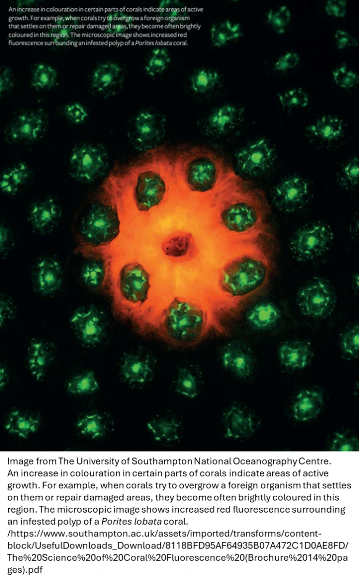
Colour intensity also increases in parts of coral that are actively growing or repairing. The choice of colours depends on the active pigments, which the coral can produce in multiple copies. This means the same species can exhibit different colours (colour morphs) depending on which genes are expressed.
|
While we know the reason for our flow cytometry samples to fluoresce, what advantage does this bring for coral? The chlorophyll is straightforward as it’s necessary for energy production, but the coral fluorescence is not fully understood although it seems to play a role in health and resilience. High light exposure is necessary for coral survival, but it does have consequences. Like our skin can get a sunburn, corals and the Zooxanthellae can become damaged by this light intensity. The fluorescent and non-fluorescent proteins of corals can act like sunblock. Soaking up the more damaging UV light and shifting it by means of fluorescence to less harmful longer wavelength light. As light is reduced deeper into the water, this fluorescence may move from a protection role to a driver of photosynthesis also.
|
|
|
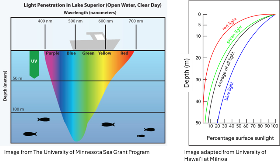
Just like in a cytometer, an excitation source is needed to make fluorescence work. However, instead of wavelength-restricted lasers, sunlight, spanning the visible and non-visible spectrum, filters through the water. As light penetrates deeper, its wavelengths become more restricted. You can almost think of water depth as a proxy for different lasers. Near the surface, you get something like a five-laser Aurora, with UV through to red light. But over 50% of visible light energy is absorbed within the first 10 meters. By 100 meters, only dim blue-green light remains, akin to little blue laser only GFP cytometer.
|
Zooxanthellae need intense light to photosynthesize, so coral is limited to shallow waters. Shallow-water reefs are bright and colorful, while deeper corals tend to appear grayish.
|
Just like how our flow cytometry experiments are best performed in a buffer free of media and other particulates, coral reefs thrive best in clear nutrient-depleted water. This clarity maximizes sunlight reaching the polyps. Turbidity in the water would reduce this light exposure, impacting the reef’s health.
|
|
|
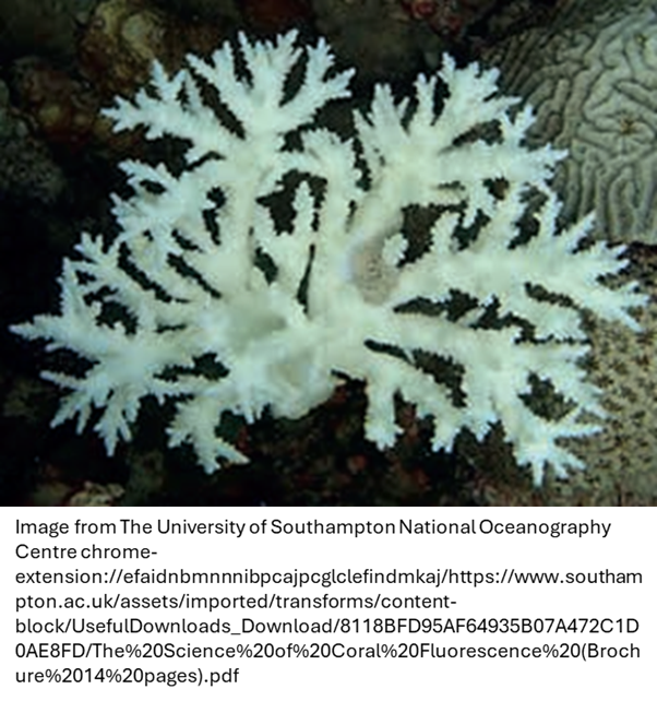
Anyone who’s had bleach come into contact with their samples knows it’s not good for them. Corals have their own version of bleaching to contend with. High temperatures or toxic water stress the symbiotic relationship between coral and zooxanthellae, causing the latter to leave. The result is the exposed hard, white skeleton of the coral, a phenomenon known as coral bleaching. This is a sign of poor reef health and has increased with the effects of climate change.
|
|
Is it necessary to think about corals during your next flow cytometry experiment Probably not. But it’s not the worst idea. After all, the 2008 Nobel Prize in Chemistry was awarded for the discovery and development of GFP, and the 2014 Nobel Prize for Chemistry was awarded for the development of super-resolved fluorescence microscopy, a technique that relied on a photoswitching protein found in coral. While I usually invite you to ask questions on the topic at the end of the newsletter, this time, those questions might be better suited for a marine biologist, but for anything flow cytometry related feel free to reach out to the staff at the FCF.
|
|
|
|
|