|
|

|
|
|
|
|
|
|
Facility News
April has to be the coolest month of the year, we got longer and warmer days, nature's healing and we have a long Easter Weekend 🐰! What else could you ask for ? Hum, I give you chocolate egg extravaganza ! As usual, I will be hiding chocolate eggs across both facilities for Easter. So on your way back from the long Easter weekend, make sure to look for them and may the best hunter WIN !
|
|
In this month FACS Tips, we're exploring the possibilities brought by the next era in flow cytometry : Image-based cell sorting. Kevin walk you through the steps required to understand this new type of dataset and it will inspire you to start a new project !
|
|
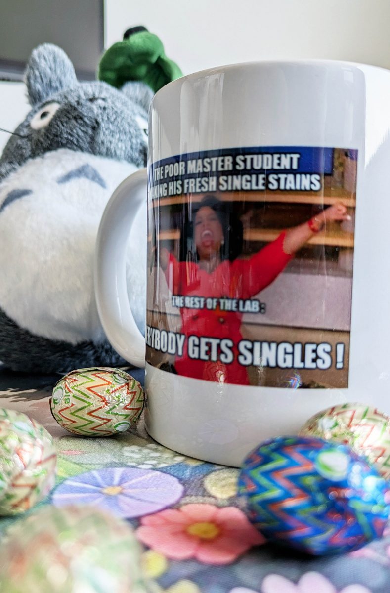
Riccardo Boccaletti won the mug this month, Congratulations to you !
|
|
|
Each month, we will ask you 3 questions about the newsletter topic. If you win, you can enter the lottery to win a unique mug designed by the FCF team !
Please take few minutes to answer the quiz HERE.
|
|
|
|
FACS Tips
|
Beyond Fluorescence: A New Era of Image-Based Cell Sorting
|
One of the promises of our new S8 sorter was that sorting decisions could be made off more than just the conventional fluorescence captured in the machine, but also by image based properties. These properties include localized fluorescence, morphological changes, marker colocalization, internalization, and more.
|
This is an exciting and promising advancement in sorting technology, enabling capabilities that were previously impossible, at least not at the high speeds achievable with the S8.
|
One challenge in implementing image-based sorting is determining the correct parameters to identify cells of interest. The S8 adds 96 imaging parameter options beyond traditional fluorescence by incorporating light loss features and three additional fluorescent channels. This creates a more complex but powerful sorting process. If you're unfamiliar with how these parameters function, it can be difficult to troubleshoot the best approach for isolating your target cells. Thankfully, in addition to the hardware tools integrated into the S8 to achieve this sorting, there have also been some additional software tools to help optimize this process.
|
|
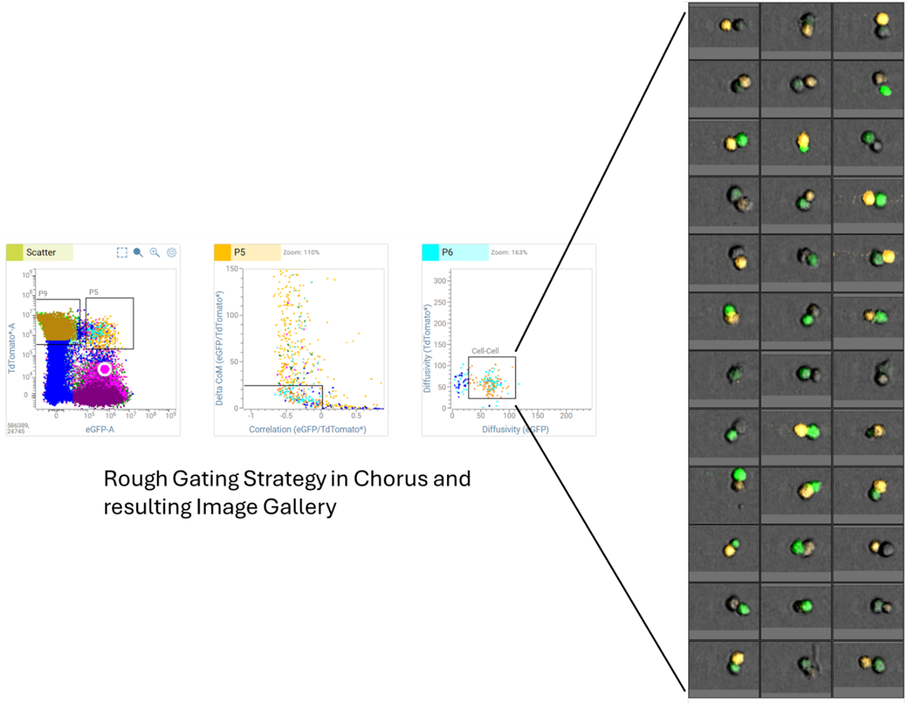
Below, we walk through an example of how we refined a gating strategy, starting with broad gating for cell-to-cell interactions using TdTomato and GFP-expressing cells and progressing to an optimized approach using image-derived parameters. Thanks to Hilma Van der Host for offering her data for this example.
|
|
|
|
In this example, we sought to identify TdTomato-expressing cells interacting with GFP-expressing cells. We began by applying a large light loss area discrimination (analogous to FSC x SSC) and gating for events positive for both markers. From there, we used several imaging parameters to filter out non-interacting cells in close proximity, as well as single cells that had non-specifically picked up both markers. This initial approach allowed us to efficiently isolate a rough population of interest for image recording and further analysis.
|
|
|
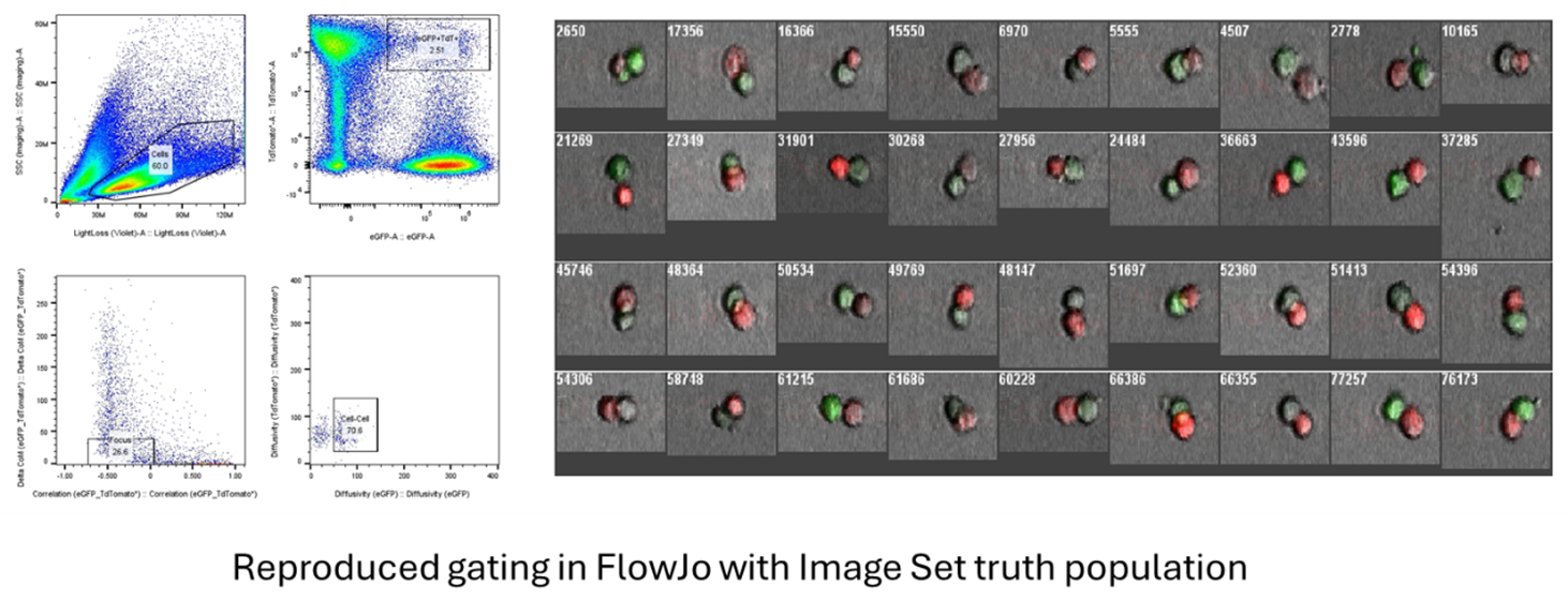
After recording 200,000 events with images, we exported the FCS files and images and used FlowJo plugins to refine our gating. First, we recreated the initial gating from Chorus in FlowJo to isolate the rough population of interest. Then, we used the CellView Lens plugin to visualize the captured images and link them to their positions on a dot plot. This plugin also provides image display controls to refine visibility.
|
|
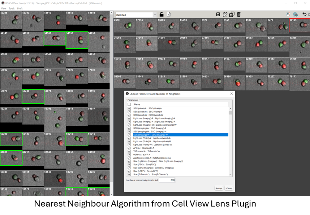
From here we can create what's called an Image Set to define a truth population of images that we think best fit the population of interest. 25 to 50 images is a good start but the more the better. If we think we’ve been too selective with our gating and have lost too many of your cells we could adjust accordingly by revisiting earlier populations in the hierarchy. If we wanted to, we could even try and just find the cells of interest from the images entirely without any gating but just by looking at the images from the CellView plugin from the start; however that will certainly take much longer.
|
|
|
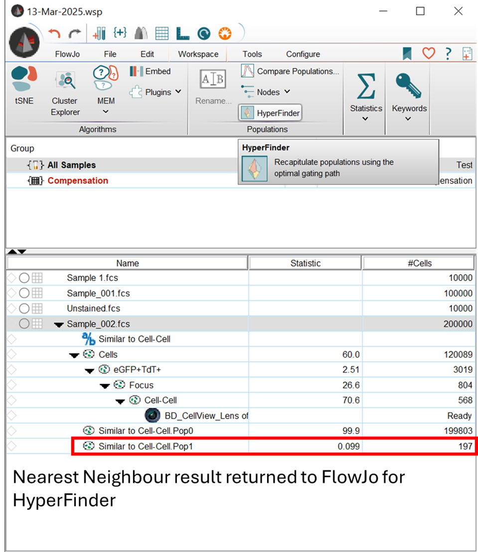
Once the Image Set was established, we applied the Nearest Neighbors algorithm to identify a predetermined number of cells that best matched our defined criteria. Here all the imaging parameters have been used to select 200 cells matching the desired cell-cell interaction we were looking for.
|
|
After having processed these images and fine tuning to ensure you have our population of interest, we can move this nearest neighbour population of cells from the Image Set back into FlowJo as a subgate of the main population.
|
|
|
|
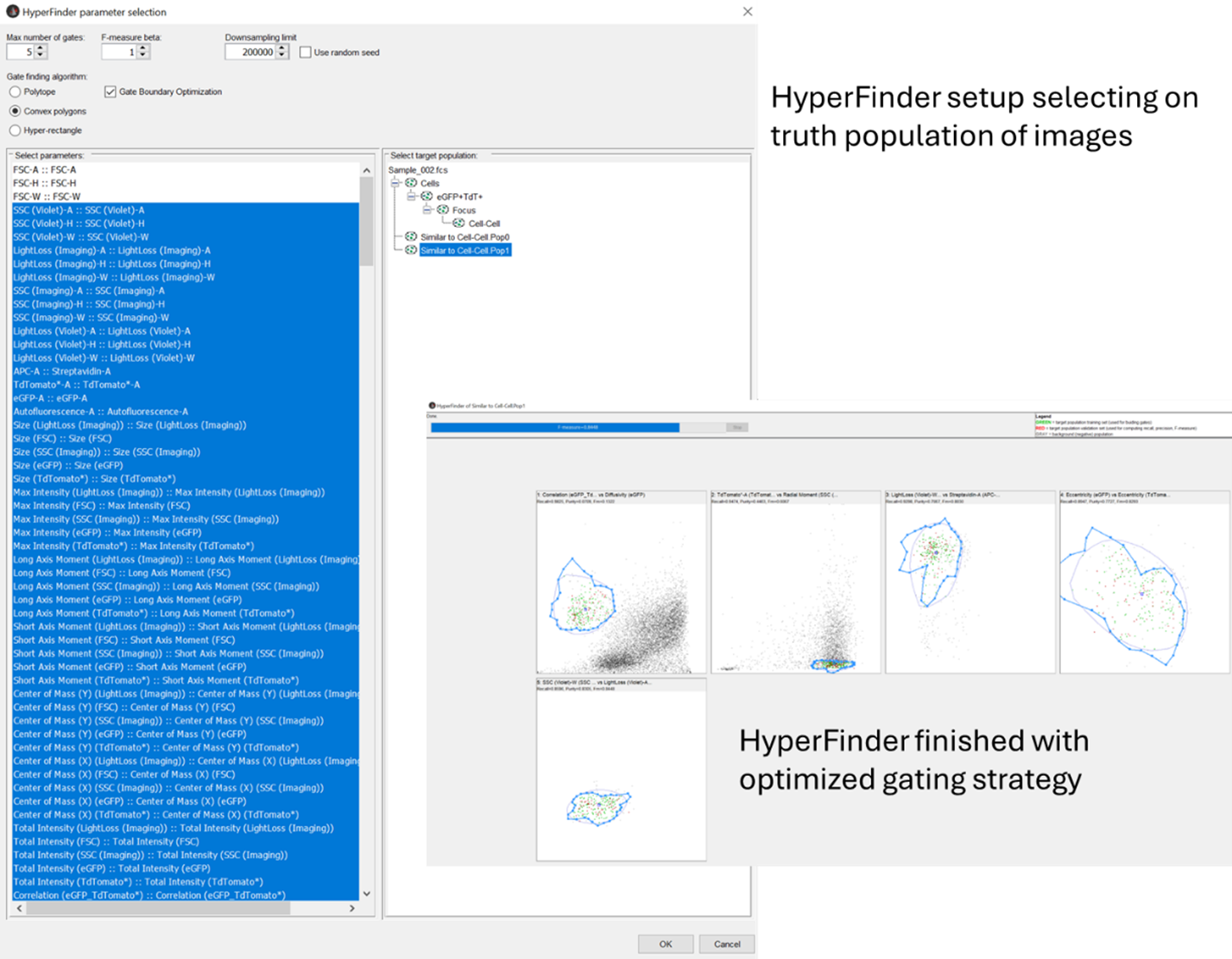
We now have the population of cells we want, we just need a gating strategy to find them. This is where we turn to HyperFinder, a new tool in FlowJo exactly for this purpose. If we click on the sample in FlowJo, and then the HyperFinder button in the Workspace Tab. By selecting your desired parameters, again here only using the imaging parameters, and hitting run, the program will find the optimized path to gate our cells in a way that can enable sorting, and give it an F score from 0 to 1 with 1 being the best.
|
|
|
As we can see this got a score of 0.8448, and now we have a more traditional 2D gating strategy to identify the cells we best selected from their imaging features.
|
To sort this population, Chorus has added a feature that now allows us to import gating from FlowJo back into the workspace and then use that as the sorting population. And there you have it, we’ve started with a rough idea of an image-based population we wanted to look at, and with a small amount of manual processing and the help of a few algorithms and we have an optimized approach. This isn’t just limited to image analysis as well, if you’ve done some dimension reduction and clustering of your data, a tSNE for example, it’s possible to gate it and import that cluster back into Chorus using a similar pathway with HyperFinder to gate and sort it.
|
Certainly, anytime we add a vast number of additional data and parameters to an analysis it will get more complicated, and while these tools do help greatly with overcoming some of these hurdles, good experiment planning at the start for what we’re looking for and how you anticipate markers/morphology to change help greatly.
|
|
If you think something like this could help with your future experiments or if you have any additional questions, make sure to reach out to the FCF team for support.
|
|
|
|
|