|
|

|
|
|
|
|
Facility News
I hope you managed to go through this winter relatively unaffected by the waves of flu and bronchitis ! Spring 🌼 is upon us and soon we will revert back to actually having sunlight after work ! Let's forget the cold ❄️ and embrace the warm weather coming to us ☀️ !
|
|
|
|
In this month FACS Tips, we talk about how the size of your fluorochomes can affect your experiments and which considerations to take to limit its impact on your data.
|
|
Nagham Alouche won the mug this month, Congratulations to you !
|
|

|
Each month, we will ask you 3 questions about the newsletter topic. If you win, you can enter the lottery to win a unique mug designed by the FCF team !
Please take few minutes to answer the quiz HERE.
|
|
|
|
FACS Tips
|
What’s Your FluoroScope
|
When selecting fluorochromes, we often focus on excitation and emission properties, spectral overlap, marker availability, and expression patterns. These factors are fundamental to panel design and can determine the success or failure of an experiment. Although one element that is almost completely forgotten is the size of the fluorochromes we choose. Fluorochrome size can influence marker performance in our experiment, yet, even across the literature, it's a rarely discussed topic.
|
|
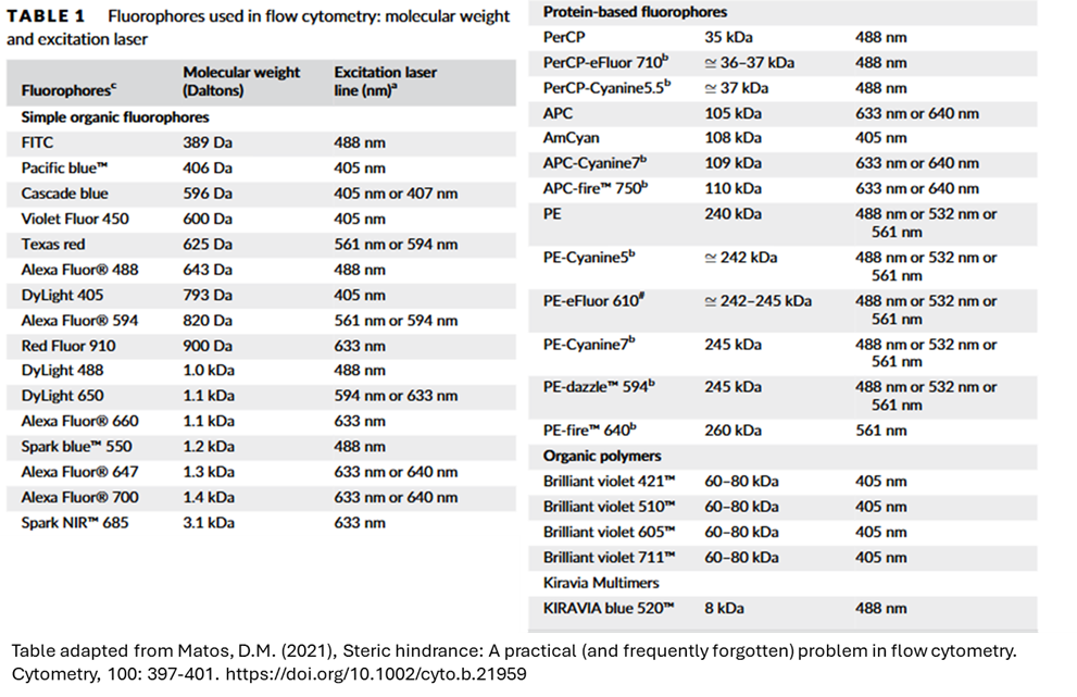
Below is a table from the paper Steric Hindrance: A Practical (and Frequently Forgotten) Problem in Flow Cytometry by Daniel Mazza Matos, MD, PhD, which lists the molecular weight (in Daltons) of commonly used fluorochromes. The size variation is striking, ranging from FITC (389 Da) to PE-Dazzle (245,000 Da). But what are the practical implications of these differences?
|
|
|
|
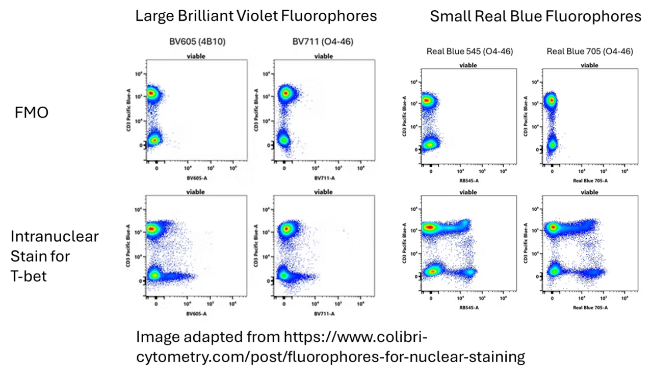
Fluorochrome size can influence staining in multiple ways. For starters, if intracellular staining is your goal, the selected fluorochromes need to get access to the site for binding their intracellular epitopes. Larger fluorochromes may struggle to navigate the permeabilized membrane compared to smaller ones. For this reason, using a large fluorochrome such as BV711 (60–80 kDa) for intracellular staining is potentially less suitable. A smaller fluorochrome would be a better choice. This is the same advice suggested in a great blog post on nuclear staining on the Colibri Cytometry website (https://www.colibri-cytometry.com/post/fluorophores-for-nuclear-staining). Additionally, it's mentioned in this blog post that nonspecific staining (background) can increase with very large fluorophore markers as they have more difficulty washing out of permeabilized cells.
|
|
|
While a more comprehensive evaluation of various molecular weight markers for intracellular staining would be a massive help in this area as a reference, it doesn’t currently exist, which is to say that at the end of the day, you need to test and validate various marker-fluorophore combinations to see what works for your experiment. However, it would certainly be advisable to go with smaller brighter dyes for intracellular/intranuclear staining. It is also important to point out that different permeabilization buffers such as Saponin and Triton generate holes in the membrane with different results and this too can play a role. For instance, if an intranuclear stain is desired it may require a more aggressive permeabilization buffer to get the desired effect.
|
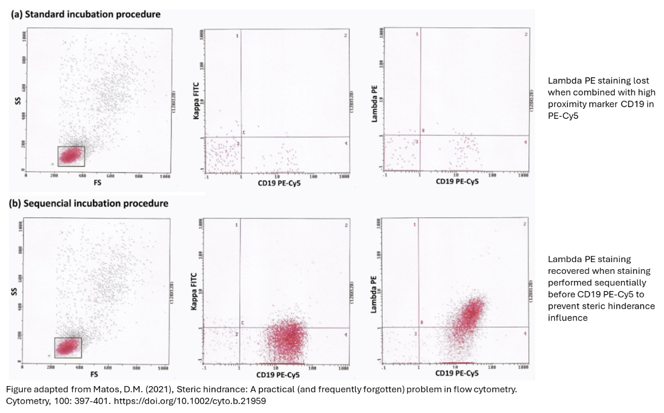
Beyond just entering the cell, fluorochrome size can have other influences in terms of binding site and proximity to other markers. In an interesting example put forward in their paper, Matos observed an issue with resolving kappa (FITC) and lambda (PE) staining in a B cell clinical assay. After troubleshooting, they suspected steric hindrance from the CD19 PE-Cy5 antibody in their panel. Steric hindrance is the effect of one monoclonal antibody disrupting the ability of another to bind to its respective target, when both of these targets appear on the same macromolecular complex. As both PE (240kDa) and PE-Cy5 (~242kDa) are heavy fluorochromes it is suspected they could more greatly inhibit one another’s ability to bind in close proximity.
|
|
To test this, they performed the same staining but in sequential incubations finishing with the CD19 stain, and they were able to recover the expression of the lambda signal in their sample. The genius of this method is by staggering the staining process they could see if just by giving the lambda staining a chance to bind first if it could do so successfully, meaning the antibody was indeed working.
|
|
|
|
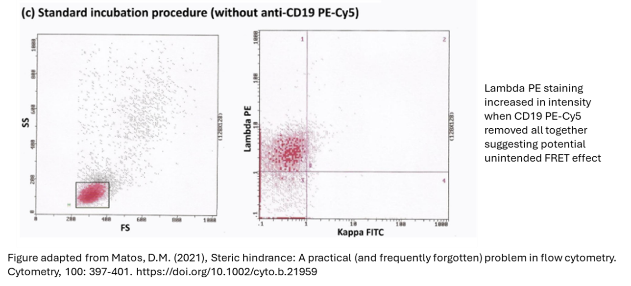
An additional realization the author had was that the lambda staining appeared weaker than anticipated. They rationalized that this could be due to some unintentional fluorescence resonance energy transfer (FRET) taking place between the Lambda PE stain and the CD19 PE-Cy5 tandem dye. Due to the proximity of these two markers, some of the desired emission signaling for quantification is instead captured by the tandem dye and acquired and counted as PE-Cy5 stain and not the PE alone. When they removed the PE-Cy5 stain all together they did in fact see the staining intensity increase for PE.
|
|
|
It’s important to point out though, as the author does in their paper, that this was a rare phenomenon, and didn’t happen with the same staining in other samples they ran. And the rare asymmetrical nature of this phenomenon makes it particularly hard to control for.
|
|
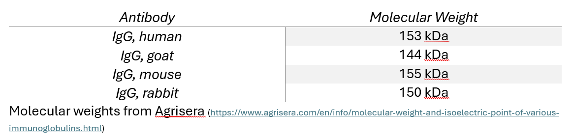
Lastly, I will mention that these fluorochromes are just part of the staining complex also including the antibody, which have their own weights. IgG and its subclasses are the predominant type used in flow cytometry staining and they have a weight of around 150kDa. This means that while a IgG rabbit FITC conjugate is only slightly heavier than the antibody alone, when conjugated instead to PE the total complex weight is x2.6 the antibody alone.
|
|
|
|
The fact that this isn’t a heavily discussed topic in panel design would lead me to align with the prevailing consensus that this is not as large a priority as ensuring the consistency of reagents and minimizing colour compensation issues. However, it does need to be something present in mind when it comes to troubleshooting. If a marker isn’t doing what you anticipated it can be an added tool in your arsenal to assess for size, hindrance, or unanticipated FRET influence on your staining. As always, the FCF team is available to answer questions and provide support.
|
|
|
|
|