|
|

|
|
|
|
|
Facility News
The finale streak of 2024 is upon us. In a bit less than 3 weeks, it will be time for - hopefully - snow ❄️, naps next to the chimney and humoungous dinners with family ! We will support you to wrap up your last experiments before the break !
|
Happy Holidays to all ⭐🌟⭐!
|
|
|
|
As in previous years the Flow Cytometry Facility (Biopole 3 and Agora/CHUV labs) will be officially closed from 6.00pm Friday December 20th 2024 through to 12.00 noon on Monday 6th January 2025. During this time there will be no FCF support or training, and no possibility to organize repairs as the service engineers from all companies are also on vacation until January 6th 2025.
|
There will be no sorting service at either site from 6pm Friday December 20th 2024 until 12.00 on January 6th 2025. Please plan your experiments accordingly.
|
Analytical Flow Cytometers
|
All analytical flow cytometers will be available for use by experienced users with valid access cards in both the Biopole 3 (AA31 and BA32) and the Agora/CHUV (227 and 233) labs during this time. However, no staff will be available for troubleshooting or help with setting up experiments. Please only use the machines if you really need to. Bookings should be made as usual on the IRIS booking system.
|
|
Please note that the November 2024 billing will include all FCF use up to and including December 15th 2024. The January 2025 billing will include December 16th to December 31st 2024 bookings as well as all of January 2025.
|
|
|
|
In this month FACS Tips, we will be talking about spectral cytometry and unmixing. We will adress how we can improve the unmixing by cleaning up reference controls.
|
|
Inès Di Resta won the mug this month, Congratulations to you !
|
|
Each month, we will ask you 3 questions about the newsletter topic. If you win, you can enter the lottery to win a unique mug designed by the FCF team !
Please take few minutes to answer the quiz HERE.
|
|
|
|
|
FACS Tips
|
Cleaning Up Unmixing in Spectral Cytometry
|
As we’ve come to realize, in our growing pursuit of more spectral cytometry experiments, unmixing can be quite a complicated game. And although it would be so much simpler if we could just use compensation beads alone, they increasingly appear to underperform compared to their cell counterparts. So, what are we to do when we have a complicated, heavily autofluorescent, single cell prep that we need to run. What is the best practice to get a good unmixing? I recently came across a blog post from a super helpful flow cytometry resource, Colibri Cytometry, which gave an interesting clean up procedure that may be useful for some of your more complex unmixing experiments.
|
In spectral cytometry, the unmixing process relies on clean, consistent, and ideally bright single-stain controls for each fluorophore used in an experiment. However, real-world samples come with the added complexity of inherent autofluorescence, which can introduce multiple unique spectra, distinct from the fluorophore signals we’re measuring. This is often manageable with simpler T cell samples, where autofluorescence is dim, but with more complex tissue samples, brighter and varied autofluorescent spectra can interfere with clean single stains. But why is this a problem?
|
During unmixing, the algorithm isolates each fluorophore’s unique signal within a fully stained sample by identifying unique spectral “signatures.” The more distinct each signature is, the easier it is for the system to separate them. Autofluorescence, however, can make each fluorophore’s signature more similar and thus harder to separate accurately. Essentially, BV605 + AF is more similar to BV650 + AF than BV605 is to BV650 alone.
|
|
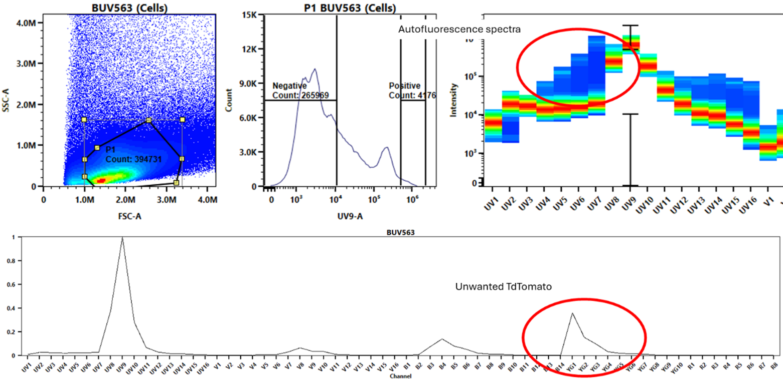
To improve unmixing in SpectroFlo Software, try gating out autofluorescence in single-stain samples, exporting this cleaner single stain as an FCS file, and re-importing it for analysis. Here’s an example using a single stain of BUV563 in highly autofluorescent liver samples, which also includes some unwanted TdTomato signal.
|
|
|
|
On initial inspection of this BUV563 single stain, the positive and negative gating histogram for unmixing may look sufficient, showing distinct populations, but hints of autofluorescence and TdTomato can still appear in the background when analyzing the spectrum.
|
|
|
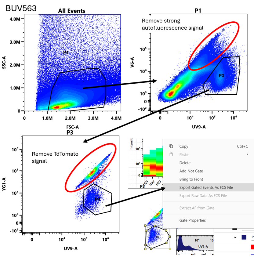
If we come back to the Raw single stain for BUV563, gate on cells (P1), then we use a combination of expected peak channel for BUV563 (detector UV9) against a common autofluorescence channel that should not overlap with the spectra of the fluorophore (V6), we can see a distinct single positive population to gate. If we then follow that down, we can remove the TdTomato in a similar fashion by again using the UV9 detector, but this time against the YG1 detector which is the peak channel for TdTomato. With a gate drawn on this single positive population, we can export these events as an FCS file that can be a standalone single positive for BUV563 without the autofluorescence background or TdTomato signal.
|
|
We can repeat this process for as many single stains as necessary that can be effectively separated from the autofluorescence background. This can get particularly tricky or unachievable if the fluorophore of interest overlaps with the autofluorescence background as they can’t be distinguished from one another on a 2D plot.
|
|
|
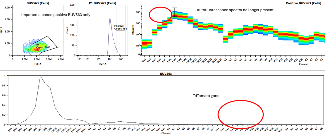
If we duplicate this experiment, we can then import the FCS file of the positive only BUV563, and then ensure to unmix with the unstained cells (assuming they’re the matching cell type and preparation as the single stain was) as the negative for this reference. As we can see, we no longer have the faint autofluorescence background signal, nor the TdTomato signal. This should make a noticeable difference in the unmixing of our fully stained samples.
|
It's also possible to use this technique to clean say GFP out of a sample to use as an unstained cell control. Take care to ensure you will have enough cells to export and that they remain a consistent and reliable sample to unmix with.
|
When it comes to problems with unmixing, your first and best solution should always be to prepare better single stains. Although this can be a time consuming and frustrating process, getting this right at the start is best for the long-term consistency of your spectral experiments. With that said, even the best made single stains can still cause unforeseen problems, so knowing these little tricks and hacks to make subtle improvements can really help with troublesome experiments.
|
|
Definitely check out the original blog on Colibri Cytometry or any of their other fantastic tips and tricks for flow cytometry. As always, feel free to reach out to the FCF staff if you have any questions.
|
|
|
|
|